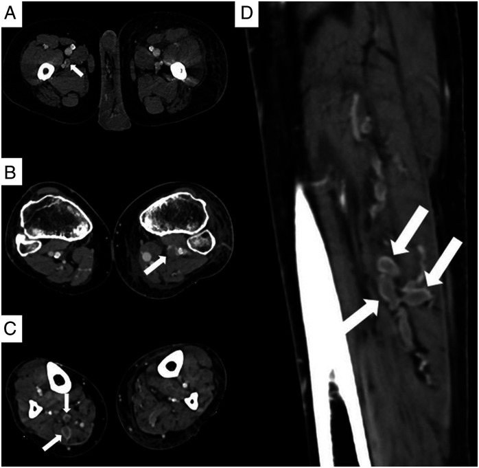
Figure 4.
(A - D): CT venogram images of three different patients. Axial (A) CT image of a 54-year-old male shows a hypodense partial thrombus in the right profunda femoris vein (arrow). Axial (D) CT image of a 61-year-old female shows hypodense filling defect in the left popliteal vein (arrow). Axial (C) and sagittal (D) images of the leg acquired in the venogram images of a 49-year-old male show expanded lumen with filling defects consistent with DVT in the muscular veins of the posterior compartment of the right leg (arrows).

