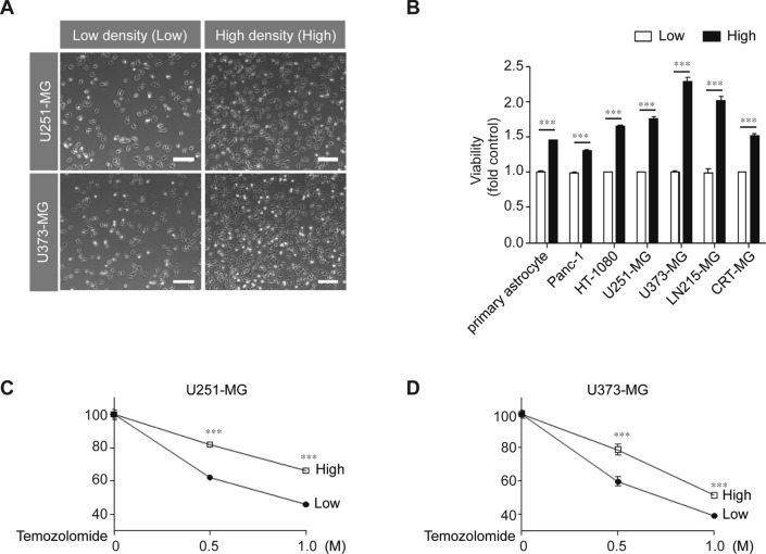
Fig. 1.
High-density culture conditions induce malignant features in various cancer cells. A U251-MG and U373-MG cells were cultured at low (9.09 × 10³ cells/cm2) or high density (2.38 × 10⁴ cells/cm2). Scale bar indicates 100 µm. B Primary astrocytes and several cancer cell lines were cultured at low or high density for 24 h, after which, cell viability was measured using a WST-1 assay (n = 3; Tukey’s post hoc test was used to detect significant differences in ANOVA, p < 0.001; asterisks indicate a significant difference compared to Low density and High density, ***p < 0.001). C, D U251-MG and U373-MG cells cultured at low or high density were treated with 0.5 M or 1 M TMZ for 24 h, after which, cell viability was measured by the WST-1 assay (n = 3; Tukey’s post hoc test was applied to detect significant differences in ANOVA, p < 0.0001; asterisks indicate a significant difference compared to 0% inhibition, ***p < 0.001).

