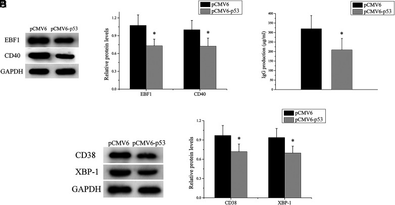
FIGURE 6.
The upregulation of p53 expression alleviates the self-reactivity of SLE B cells. (A and B) EBF1 and CD40 expression levels in SLE B cells transfected with p53 expression plasmid or negative control were analyzed by Western blot. One representative blot is shown (A). The intensity of the bands was semiquantitated and normalized to GAPDH (B). (C) SLE B cells transfected with p53 expression plasmid or negative control, where the transfected cells were cocultured with purified autologous CD4+ T cells, and IgG levels in the culture supernatants were measured by ELISA. (D and E) CD38 and XBP-1 expression levels in cocultured cells were analyzed by Western blot. One representative blot is shown (D). The intensity of the bands was semiquantitated and normalized to GAPDH (E). Data represent the mean of three independent experiments per group. *p < 0.05.

