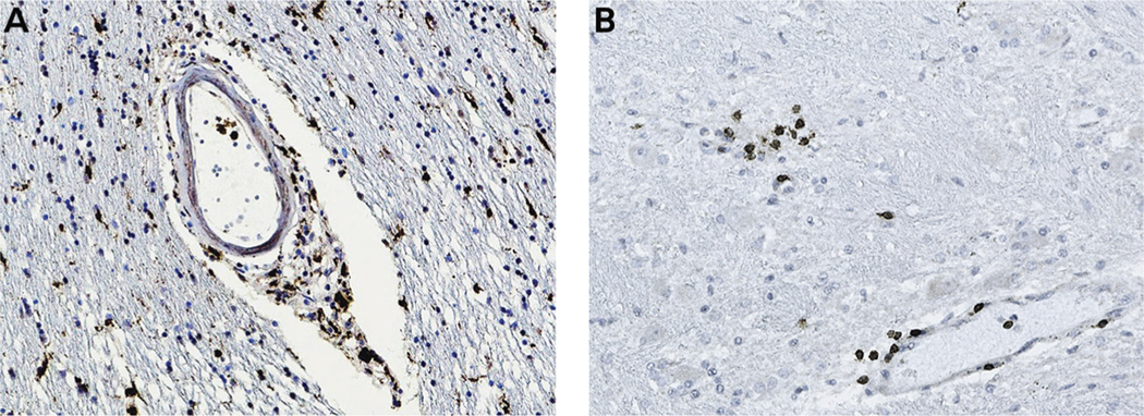
FIGURE 11-2. Central nervous system inflammation in COVID-19. Autopsy brain tissue was stained with monoclonal antibodies to immune cellular markers. Diaminobenzidine was used as a chromogen, which gives a brown-colored precipitate. Panel A shows infiltration of macrophages staining for CD68 in the perivascular region and parenchyma, and panel B shows infiltration of CD3 T cells in foci around the blood vessels and the parenchyma.
Figure courtesy of Rebecca Folkerth, MD (provided autopsy tissue) and Myounghwa Lee, PhD (performed immunostaining).

