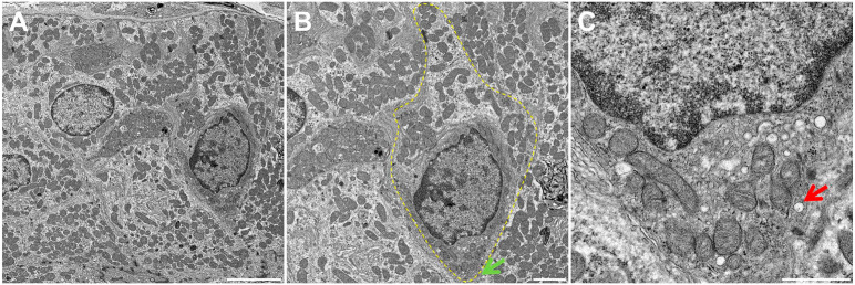
Figure 3.
Fine structure of Tuft cells (TC) of human SMG. Shown is a transmission electron micrograph of TC in a human SMG striated duct (A). Note the bottle-like cell shape at the nucleus with narrow apical and basal portions consistent with TC morphology (B). There are numerous apical vesicles in the supranuclear cytoplasm (C). The yellow dotted line indicates a TC while the green and red arrows denote characteristic apical microvilli and the tubulovesicular system. Scale bars represent 5 (A), 2 (B), and 1 (C) µm, respectively. Abbreviations: SMG, submandibular glands; TC, Tuft cells.

