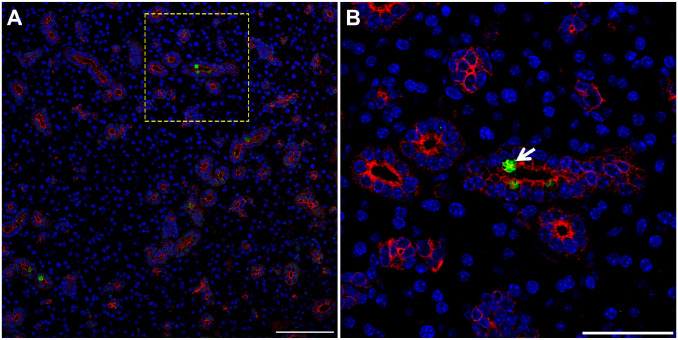
Figure 4.
TC are present in mouse SMG. Five microns of a paraffin embedded SMG section were stained with rabbit anti-POU2F3 and mouse anti-cytokeratin-7, followed by anti-rabbit Alexa Fluor 488 (green) and anti-mouse Alexa Fluor 568 IgG (red) secondary antibodies and counterstained with 4,’6-diamidino-2-phenylindole (blue). Images were analyzed using a Stellaris microscope at low (A) and high (B) magnifications. Yellow dotted lines indicate area that was magnified. White arrow denotes TC in SMG duct. Images are representative of n = 3 specimens. Scale bars from low and high magnification represent 100 and 50 µm, respectively. Abbreviations: SMG, submandibular glands; TC, Tuft cells.

