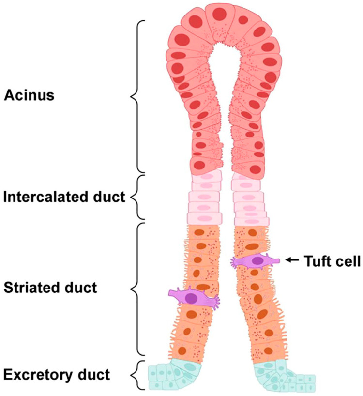
Figure 7.

Diagram representing the localization of Tufts cells in salivary glands based on the findings obtained in this study. Salivary gland structure consists of secretory acini and ducts (i.e., intercalated, striated and excretory). In this diagram, Tuft cells were depicted by their distinct bottle-shaped morphology and well-developed “tuft-like” microvilli at the apical surface. As for their location, these cells were restricted to striated ductal epithelia. Created using Biorender.com.
