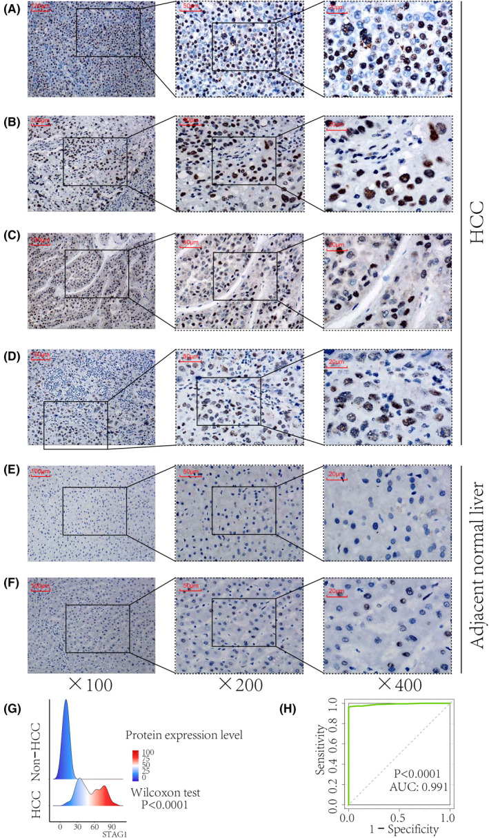
Fig. 4.

In‐house immunohistochemistry staining of STAG1 transcriptional factor in HCC tissue samples. STAG1 protein was stained in the nucleus of the HCC cells (A–D) and compared with healthy cells (E–F). Increased protein expression of STAG1 in HCC displayed a strong ability in differentiating HCC from non‐HCC tissue samples (G–H). HCC, hepatocellular carcinoma. The scale bars in panels A–F represent 100, 50, and 20 μm, from left to right.
