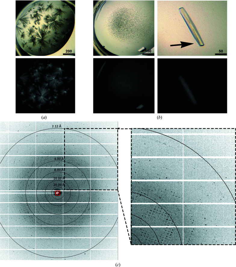
Figure 4.
(a, b) Images of PorXFJ crystals. The upper images were taken in visible light, while the lower images were taken in UV light, demonstrating that the crystals are indeed protein crystals. (a) Original crystallization condition. Scale bars are labeled in µm. (b) Optimized crystals after successive rounds of seeding. The arrow indicated the area that diffraction data were collected from. (c) Representative diffraction image.

