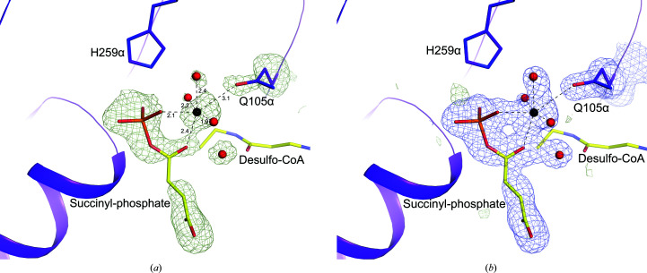
Figure 3.
Binding of succinyl-phosphate to E105αQ GTPSCS. The maps show the binding of succinyl-phosphate, a magnesium ion, water molecules and the side chain of Gln105α. Succinyl-phosphate and part of desulfo-CoA are shown as sticks with yellow C atoms. The magnesium ion and water molecules are shown as black and red spheres, with ionic interactions represented by dashed lines. Portions of the α-subunit are represented by a purple ribbon diagram with the side chain of Gln105α and the active-site histidine, His259α, shown as sticks with purple C atoms. Other atoms are coloured according to type. (a) The omit map is contoured in light green at 3σ. Distances for the ionic interactions are given in Å. (b) The F o − F c map is contoured in light green at 3σ and the 2F o − F c map is contoured in blue at 1σ. This figure and Figs. 4 ▸, 6 ▸ and 7 ▸ were drawn using PyMOL (version 2.4.0; Schrödinger).

