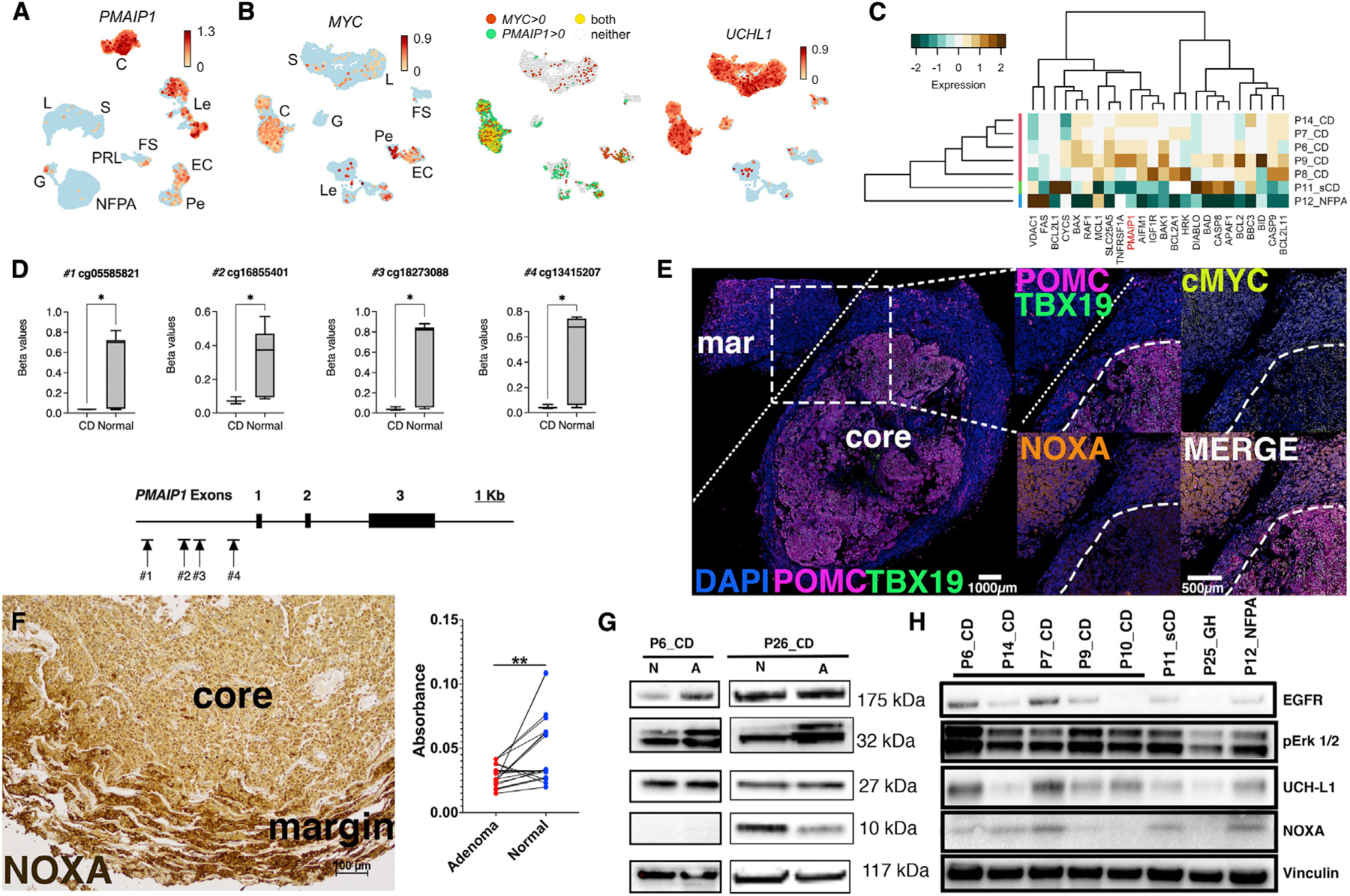
Figure 5. Human adenomas causing wild-type CD have discrepant PMAIP1 transcript and noxa abundance.

(A) UMAP plot demonstrating PMAIP1 abundance in CD adenomas (C) but not in PRL, G, or NFPA adenomas. PMAIP1 was also detected at lower levels in Les and ECs.
(B) UMAP plot showing MYC upregulation in CD corticotrophs but not other hormone-producing cells (left panel). MYC expression was also detected in Pes, ECs, and Les. Middle panel: UMAP plot showing overlap of MYC and PMAIP1 expression is mostly limited to CD corticotrophs (yellow dots). Right panel: UMAP plot showing UCHL1 abundance in most hormone-producing cell types in CD.
(C) Bulk RNA-seq of CD and non-CD samples verified overexpression of pro-apoptotic genes including PMAIP1 in CD tissues.
(D) DNA methylation levels (beta values) at CpG sites associated with the PMAIP1 promoter methylation demonstrating hypomethylation in CD (n = 3) compared with normal (autopsy-derived, n = 20) pituitary glands. *p < 0.05.
(E) Multiplex immunohistochemistry (mIHC) of 5 μm thick sections from a CD adenoma. Insets from the core-margin boundary represented by white dashed lines. White bar: 100 μm. Core-margin boundary identified by overlaying expression of POMC, TBX19, and DAPI. Compared with the margin, core adenoma cells show robust overexpression of POMC, TBX19, and MYC; however, noxa expression is decreased within the adenoma core.
(F) Representative image from noxa IHC in independent adenoma/normal pairs (n = 10). Pairwise analysis of noxa deconvoluted IHC images (absorbance = mean pixel intensity count per pixel) showing suppressed noxa signal in CD adenomas compared with normal (margin) tissues (p = 0.0013; 95% confidence interval [CI] −0.027 to −0.007). Scale bar: 100 μM.
(G) Expected epithelial growth factor (EGF) signaling upregulation and ERK1/2 phosphorylation were found in human CD adenoma primary cell lines (P6_CD and P26_CD). A, adenoma (core); N, normal (margin) pituitary gland. Noxa was undetectable or decreased in core adenomas.
(H) A survey of human primary CD adenoma cell lines revealed variable noxa expression compared with sCD adenoma, GH adenoma, and NFPA. CD, corticotroph adenoma causing Cushing’s disease; sCD, hormonally silent corticotroph adenoma; GH, growth-hormone-secreting adenoma; NFPA, non-functioning pituitary adenoma.
