
Fig. 1.
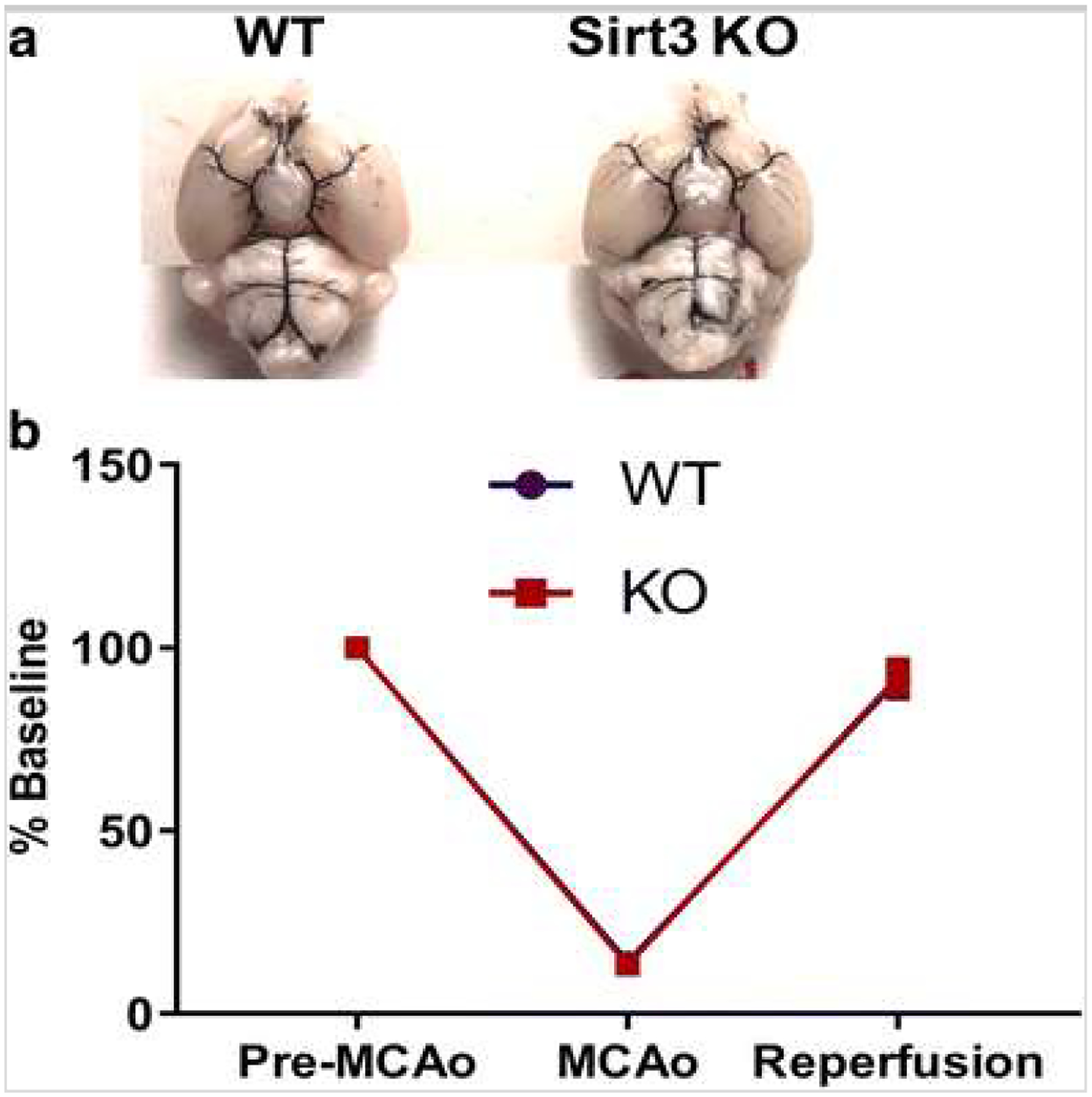
a Macroscopic analysis of the cerebral arterial vasculature shows no differences between Sirt KO and WT. Representative image of India ink staining in WT and KO mice shows branching of the internal carotid artery, anastomosis of internal carotid and basilar territories (n = 5/group). b Cortical perfusion during ischemia assessed by laser Doppler flowmetry (%LDF reduction) shows the reduction in perfusion during ischemia, and recovery of blood flow during reperfusion wag not different between genotypes