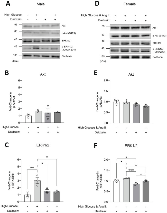
Figure 6.
Total and phosphorylated Akt and ERK1/2 expression in HPCs. (A-C) Differentiated male HPCs were treated with high glucose (20-25 mM), daidzein (10 μM), or high glucose with daidzein for 24 hours. Differentiated HPCs with 0.1% DMSO were used as controls. (A) Immunoblots of total and phospho Akt and ERK1/2 from male HPC lysates with cadherin shown as a loading control. (B & C) Densitometry from Akt (B) and ERK1/2 (C) immunoblots from 3 separate biological replicates (n = 3) normalized to control cells. (D-F) Differentiated female HPCs were treated with a combination of high glucose (25 mM)/1 µM angiotensin II (Ang II), daidzein (10 µM), or high glucose/Ang II and daidzein for 24 hours. Differentiated HPCs with 19.5 mM mannitol and 0.1% DMSO were used as controls. (D) Immunoblots of total and phospho Akt and ERK1/2 from female HPC lysates with cadherin shown as a loading control. (E & F) Densitometry from Akt (E) and ERK1/2 (F) immunoblots from 3 separate biological replicates (n = 3) normalized to control cells.
Note. All data are presented as mean ± standard error of the mean. Statistical significance compared to control cells was determined using one-way ANOVA with post-hoc Sidak’s test. Significance is marked by asterisks. HPCs = human podocyte cells; ANOVA = analysis of variance; ERK = extracellular signal-regulated kinase; DMSO = dimethyl sulfoxide.
*P < .05. **P < .01. ***P < .001.

