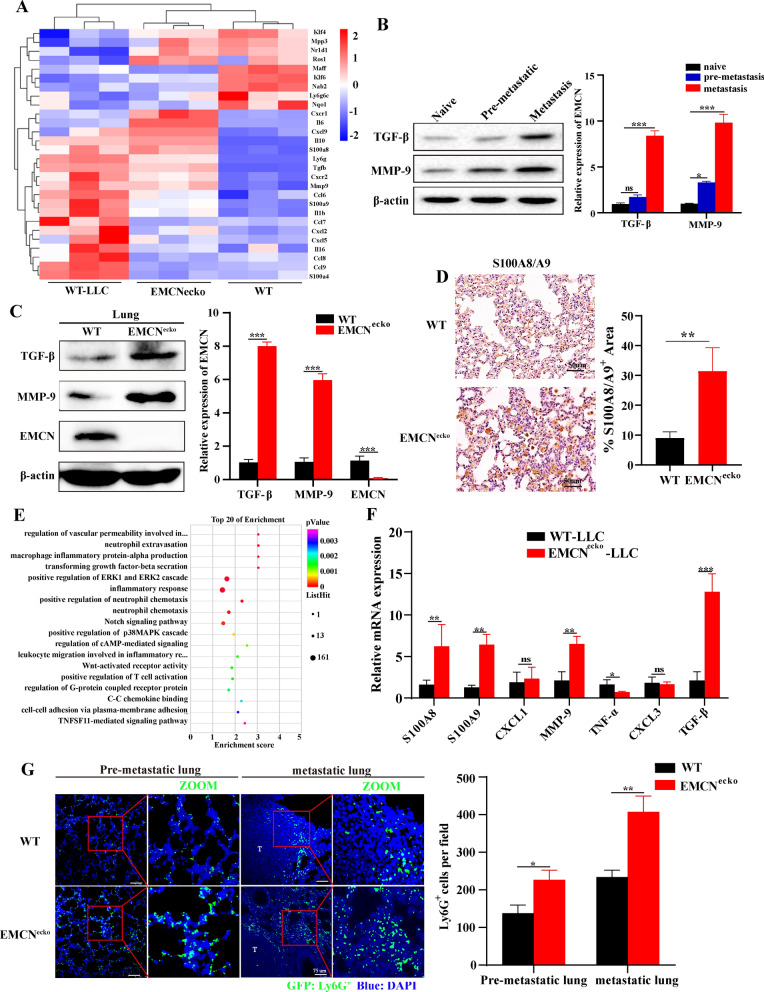
Fig. 4.
EMCN deficiency leads to the formation of a lung premetastatic niche. A Representative heatmap of differentially expressed genes in naive lung (WT), lung in the microenvironment before tumor metastasis (subcutaneous inoculation of tumor for 21 days) and EMCNecko mouse lung. Each row represents a gene, and each column represents a group of mice. B TGF-β and MMP-9 expression in naive, premetastatic and metastatic lungs. TGF-β and MMP-9 expression levels in naive, premetastatic and metastatic lungs were quantified by Western blotting. Each experiment was repeated thrice. (Data represent the mean ± SD). Statistical analysis was performed using one-way ANOVA. *p < 0.05, ***p < 0.001. C Lungs were isolated from WT and EMCNecko mice and lysed for Western blot analysis of the indicated proteins. Bands were quantified using ImageJ in F. Each experiment was repeated thrice, and the statistical significance of differences between gray intensities was determined using the unpaired Student’s t test. D S100A8/A9 staining in the lungs of WT and EMCNecko mice showed significant S100A8/A9-positive cell infiltration in the lungs of EMCNecko mice. E KEGG analyses of the significantly downregulated and upregulated genes in the lungs of WT and EMCNecko tumor-bearing mice (n = 3 biological replicates). F The mRNA levels of representative key factors were measured by quantitative PCR. RNA was extracted from the lungs of WT and EMCNecko mice inoculated with tumors (n = 3 biological replicates). G Neutrophils in the premetastatic lung (capable of reaching lungs but unable to grow up) and metastatic lung were determined by immunofluorescence staining in WT and EMCNecko mice. Scale bars, 75 μm (left). The number of Ly6G-positive cells was quantified by ImageJ (right)

