
FIGURE 2.
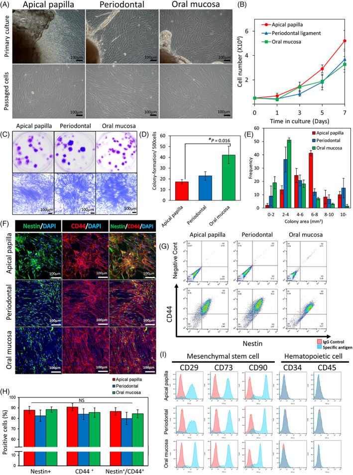
Characterization of human APDCs, PDLDCs, and OMSDCs. (A) Morphology of cultured APDCs, PDLDCs, and OMSDCs in vitro. Upper panel: primary cultured cells around the explanted tissue; lower panel: exponential growth of passaged cells in monolayer culture. (B) Cell growth curves of APDCs, PDLDCs, and OMSDCS (n = 5, patient‐matched). (C) Photographs of colony‐forming unit fibroblasts from APDCs, PDLDCs, and OMSDCs at 14 days. (D) Colony‐formation assays for APDCs, PDLDCs, and OMSDCs (n = 5, patient‐matched). Colonies stained with crystal violet. (E) Measurement of colony areas of APDCs, PDLDCs, and OMSDCs (n = 4, patient‐matched). (F) Immunohistochemistry for nestin and CD44. (G) Flow cytometry analysis of the expression of nestin and CD44. (H) Percent expression of nestin, CD44, and nestin/CD44 in cells (n = 3, patient‐matched). (I) Expression of surface markers of APDCs, PDLDCs, and OMSDCs. Average data are expressed as mean ± standard error (SE). Abbreviations: DAPI, 4′,6‐diamidino‐2‐phenylindole. *p < 0.05