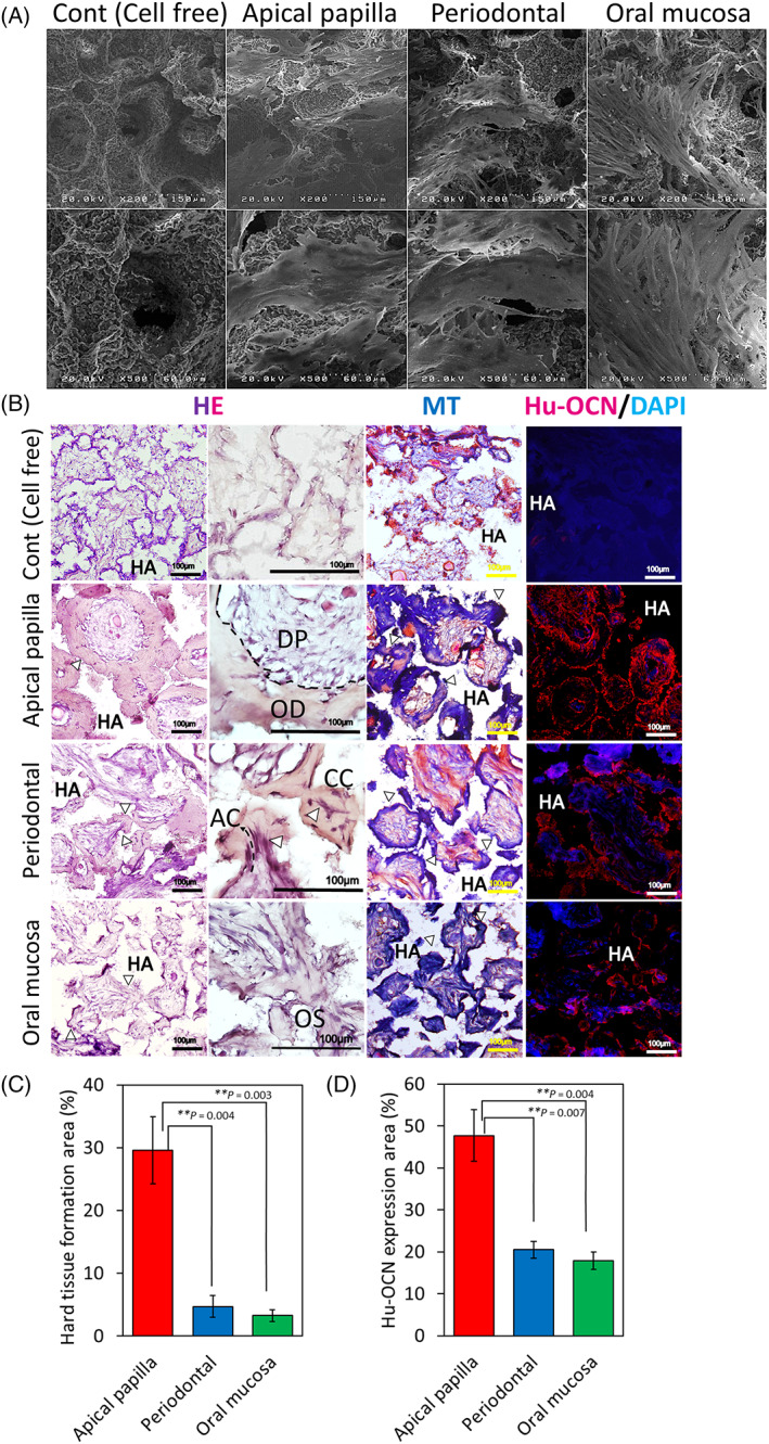
FIGURE 6.

In vivo hard tissue‐forming ability of APDCs, PDLDCS, and OMSDCs. (A) SEM images of cells adhered to porous hydroxyapatite scaffolds. (B) HE staining, Masson's trichrome staining, and immunohistochemical analysis of transplanted tissues. HE staining revealed hard/mineralized‐tissue forming ability in vivo. The generated hard tissues were dental pulp‐like and osteodentin‐like structures in APDCs, thin cellular cementum‐like or acellular cementum‐like tissues with Sharpey's fiber‐like structures (arrow head) in PDLDCs, and thin immature osteoid‐like tissues in OMSDCs. Masson's trichrome staining revealed that regenerated hard tissues were composed of collagen fibers. Immunofluorescence staining was performed for human osteocalcin. (C) Quantitative analysis of the area with hard tissue formation (n = 3, patient‐matched). (D) Quantitative analysis of human osteocalcin area (n = 3, patient‐matched). Average data are expressed as mean ± SE. Abbreviations: Cont., control group; DAPI, 4′,6‐diamidino‐2‐phenylindole; HA, hydroxyapatite; HE, hematoxylin–eosin staining; Hu‐OCN, human osteocalcin; MT, Masson's trichrome staining. Arrowhead: hard tissue. **p < 0.01
