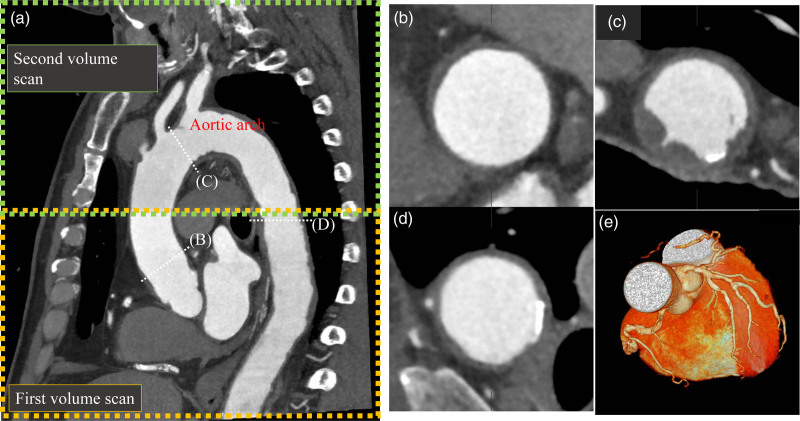
Fig. 1.
Computed tomography coronary angiography with wide-volume scanning for the aortic arch and coronary plaque imaging. (a) Oblique image of the thoracic aorta and heart. A combined image of the first and second volume scans (wide-volume scan) is automatically generated using 320-row multidetector computed tomography. (b) Aortic plaque in the ascending aorta. (c) Aortic arch plaque with protrusion. (d) Aortic plaque with calcification. (e) Volume rendering image of the coronary arteries.

