Figure 2.
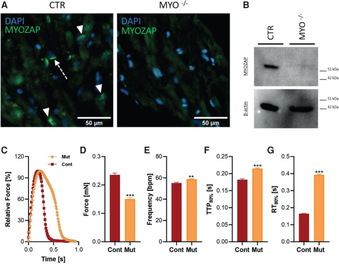
Immunohistochemical and functional studies. The absence of the MYZAP protein in proband tissue was demonstrated by catalyzed fluorescent reporter deposition immunohistochemistry and western blot (WB). (A) In the control sample (CTR), epitopes corresponding to the carboxy-terminal end of the MYZAP protein were distributed in the perinuclear cytoplasm (green signal; arrowhead) and in dense bands perpendicular to myocyte orientation (green signal; arrow). The signal was completely absent in the proband tissue (MYZAP−/−). (B) The absence of the MYZAP signal in the proband tissue was demonstrated by western blot. A β-actin signal on the same membrane was used for internal control (total protein loading control is provided in Supplemental Fig. S1). The bottom panels contain the results of the functional characterization of the engineered heart tissues (EHTs) of patient-derived human induced pluripotent stem cell cardiomyocytes (hiPSC-CMs). (C) The average contraction peaks of EHTs in 1.8 mM Ca2+ Tyrode's solution under 1-Hz pacing at 37°C. (D–G) Functional parameters of absolute force (D), frequency (beats per minute, bpm) (E), time to peak 80% (TTP80%, F), and relaxation time (RT80%, G) measured in 1.8 mM Ca2+ Tyrode's solution under 1-Hz pacing at 37 °C. n = 8 patient EHTs and 11 control EHTs. Statistical calculations were carried out by a two-tailed Student's t-test. (**) P < 0.01, (***) P < 0.001.

