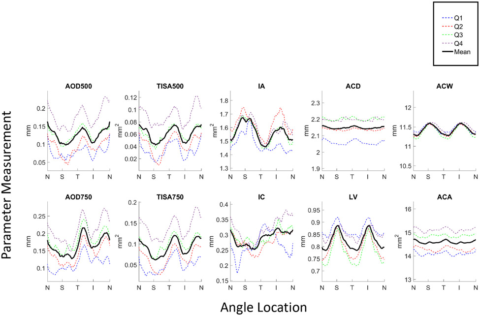
Figure 1.
Angle parameter measurements averaged over 32 images plotted by angle location (N = nasal, S = superior, T = temporal, I = inferior) for each of the 4 quartiles, Q1 (narrowest angles; blue dotted line) through Q4 (widest angles; purple dotted line), and averaged across all quartiles (black solid line).

