. 2022 Oct 3;11:e79128. doi: 10.7554/eLife.79128

Figure 2. S6K1 and Iso-2 bind to DNA damage/repair proteins.

(a) Venn diagram representing DNA damage response/repair proteins that bound specifically to HT-S6K1 isoforms in transfected HEK293 cells. Proteins marked in blue were validated by reciprocal immunoprecipitations. (b,c) Western blot of total lysate (L) and flow through (FT) of Halo-tag pulldown from HEK293 cells transfected with either Halo-tag expressing control vector (HT), HT-S6K1 (S6K1) or HT-Iso-2 (Iso-2) (b, left panel and c, upper panel). Western blot of Halo-tag pulldown from the same cells (b, right panel c, lower panel). (d) Western blot of total lysate (L) and flow through (FT) of Halo-tag pulldown from HeLa cells transfected with either Halo-tag expressing control vector (HT), HT-S6K1 (S6K1) or HT-Iso-2 (Iso-2) (upper panel). Western blot of Halo-tag pulldown from the same cells transfected with either Halo-tag expressing control vector (HT), S6K1 or Iso-2 (lower panel). (e) Western blot of total lysate from HEK293 cells co-transfected with either Halo-tag expressing control vector (HT), HT-S6K1 (S6K1) or HT-Iso-2 (Iso-2) and HA-Cdk1 (upper panel). Western blot of immunoprecipitation of lysates of these cells with anti-HA antibody (lower panel). (f). Western blot analysis of HEK293 cells transfected as described in b (left panel). Western blot of immunoprecipitation of lysates of these cells with anti-PCNA antibody (right panel).

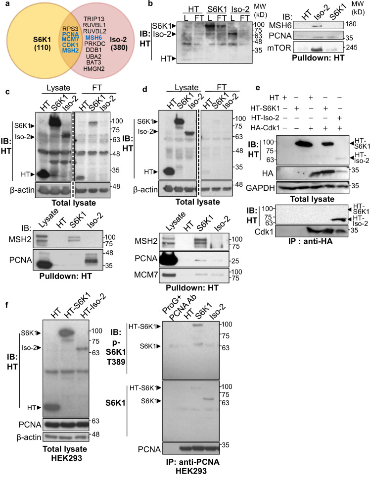
Figure 2—source data 1. Western blots of Figure 2.

Figure 2.

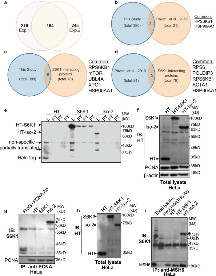
Figure 2—figure supplement 1. S6K1 isoforms interact with proteins involved in DNA repair.

Figure 2—figure supplement 1.

(a) Venn diagram representing the overlap of proteins identified in two independent proteomic analyses. Proteins bound to HT-S6K1 isoforms in HEK293 cells transfected with either HT, HT-S6K1, or HT-Iso-2 from Halo-tag pulldown assay were analyzed by Mass-spectrometry. The numbers represent the number of proteins which specifically bound to either of the HT-S6K1 isoforms and not to the HT control. Exp. 1 was performed using the conventional proteomic method (gel extracted samples) and Exp. 2 was performed using the ‘on bead digestion’ method. (b) Venn diagram representing the overlap of S6K1 interactors identified in this study (both S6K1 and Iso-2) and those reported in Pavan, et al. 2016 (Raught et al., 2004). Only high-scoring S6K1 interactions with SAINT probability (SP) >0.9 and/or containing potential phosphorylation site (RSRXXT/S) were used for comparison. (c) Venn diagram representing the overlap of S6K1 interactors identified in this study and BioGrid (Biological General Repository for Interaction Datasets https://thebiogrid.org/2). (d) Venn diagram representing the overlap of high-scoring S6K1 interactors identified in the Pavan study compared to BioGrid. (e) Western blot of total lysates (L) and flow through (FT) of HEK293 cells described used for proteomic analysis in Exp. 2 (triplicates). (f,g) HeLa cells transfected as described above were used for immunoprecipitation using PCNA antibody. Western blot analysis of total lysates (f) and immunoprecipitated proteins (g). (h,i) HeLa cells transfected as described above were used for immunoprecipitation using MSH6 antibody. Western blot analysis of total lysates (h) and immunoprecipitated proteins (i).
Figure 2—figure supplement 1—source data 1. Western blots of Figure 2—figure supplement 1.
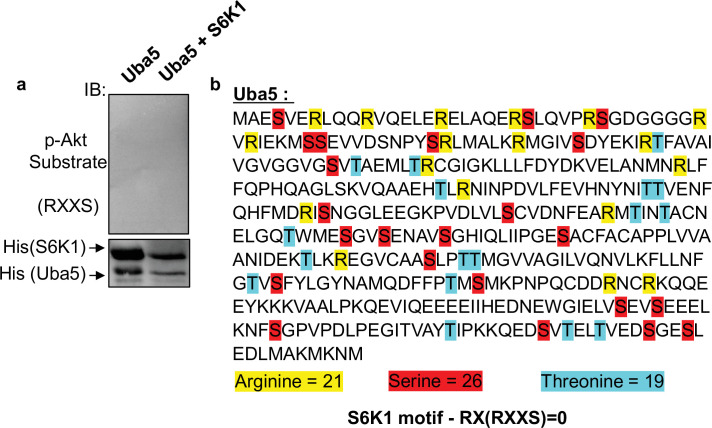
Figure 2—figure supplement 2. S6K1 does not phosphorylate unrelated protein Uba5.

Figure 2—figure supplement 2.

(a) Non-radioactive in vitro kinase assay using recombinant His-S6K1 [T412E] and recombinant Uba5 (gift from the laboratory of Dr. Reuven Weiner). Reaction products were resolved by SDS-PAGE and analyzed by Western Blot using an antibody against the S6K1 phosphorylation motif (p-Akt substrate, RXXS) or an antibody to detect the recombinant proteins. (b) Protein sequence of Uba5. Arginines, serines and threonines are highlighted showing absence of an RXXS motif.