Figure 1.
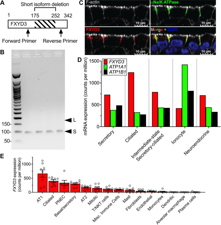
FXYD3 is expressed in human airway epithelial cells. A: primer design for detecting short vs. long FXYD3 isoforms. B: agarose gel of PCR products is shown for PCR reactions (35 cycles) containing cDNA obtained from well-differentiated airway epithelia (n = 4 donors); L, long-FXYD3; S, short-FXYD3. C: FXYD3 colocalizes with the α subunit of Na/K ATPase in the basolateral membrane of airway epithelia. Representative image from experiments performed on 5 donors. D: summary of single-cell RNA sequencing data of an airway epithelium cultured from cartilaginous airways. Bars represent expression level from pooled cells obtained from 5 cultures from 1 biological replicate, thus standard deviations are not shown. E: summary of previously published (15) single-cell RNA sequencing data of human lung performed on 8 human donors. Each dot represents 1 human donor and error bars represent the standard error of the mean. Mean counts per million ± SD; AT1 660.0 ± 369.3, Ciliated 403.2 ± 137.0, PNEC 336.8 ± 216.4, basal/secretory 302.0 ± 98.99, AT1 177.3 ± 65.92, mitotic 150.3 ± 112.7, T/NKT cells 112.5 ± 70.63, misc. immune cells 98.21 ± 32.75, mast 78.91 ± 82.96, fibroblasts 48.11 ± 45.61, endothelial 37.59 ± 20.93, monocytes 23.34 ± 44.66, dendritic 6.13 ± 5.14, alveolar macrophage 4.29 ± 2.22, plasma cells 6.48 ± 14.42. P values were calculated using a one-way ANOVA with Tukey correction and are presented in Table 4 for clarity. NKT, natural kill T; PNEC, pulmonary neuroendocrine cell.

