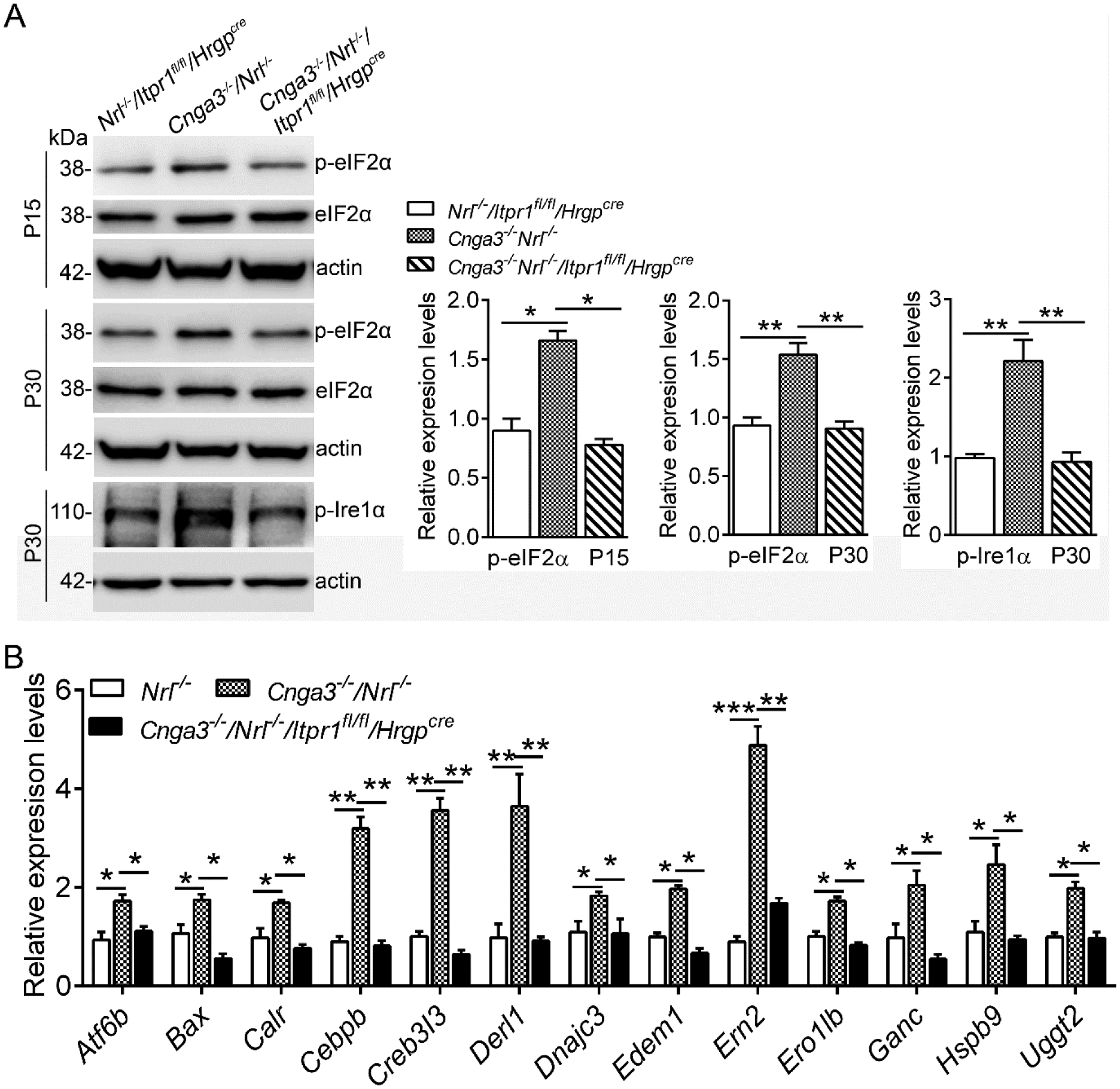
Figure 1. Deletion of IP3R1 reduced UPR/ER stress in CNG channel-deficient retinas.

A. Expression levels of ER stress marker proteins in mouse retinas at P15 and P30 were analyzed by immunoblotting. Shown are representative immunoblotting images of these detections and corresponding quantitative analysis, following normalization to internal loading control β-actin. B. Expression of UPR genes was evaluated in mouse retinas at P15 by PCR array. Shown are PCR array results. Data are presented as mean ± SEM of 3–4 independent assays using retinas from 8–10 mice (*p < 0.05, **p < 0.01, ***p < 0.001).
