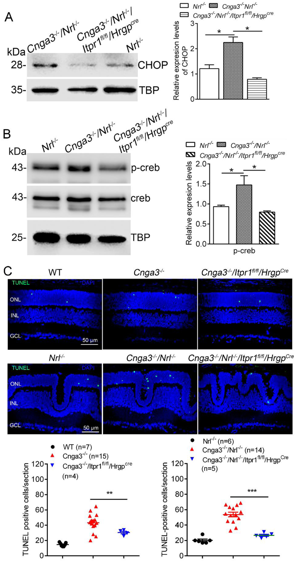
Figure 2. Deletion of IP3R1 reduced ER stress downstream signaling and apoptosis in CNG channel-deficient retinas.

A-B. Expression of CHOP and p-Creb were evaluated in mouse retinas by immunoblotting. Shown are representative immunoblotting images for CHOP at P15 (A) and p-Creb at P30 (B) and the corresponding quantitative analysis. C. Photoreceptor apoptosis was evaluated by TUNEL labeling on retinal sections of CNG channel-deficient mice at P15. Shown are representative confocal images of TUNEL labeling and correlating quantitative analysis. ONL, outer nuclear layer; INL, inner nuclear layer. Data are presented as mean ± SEM of 3–4 independent assays using retinas/eye sections from 4–10 mice (*p < 0.05, **p < 0.01, ***p < 0.001).
