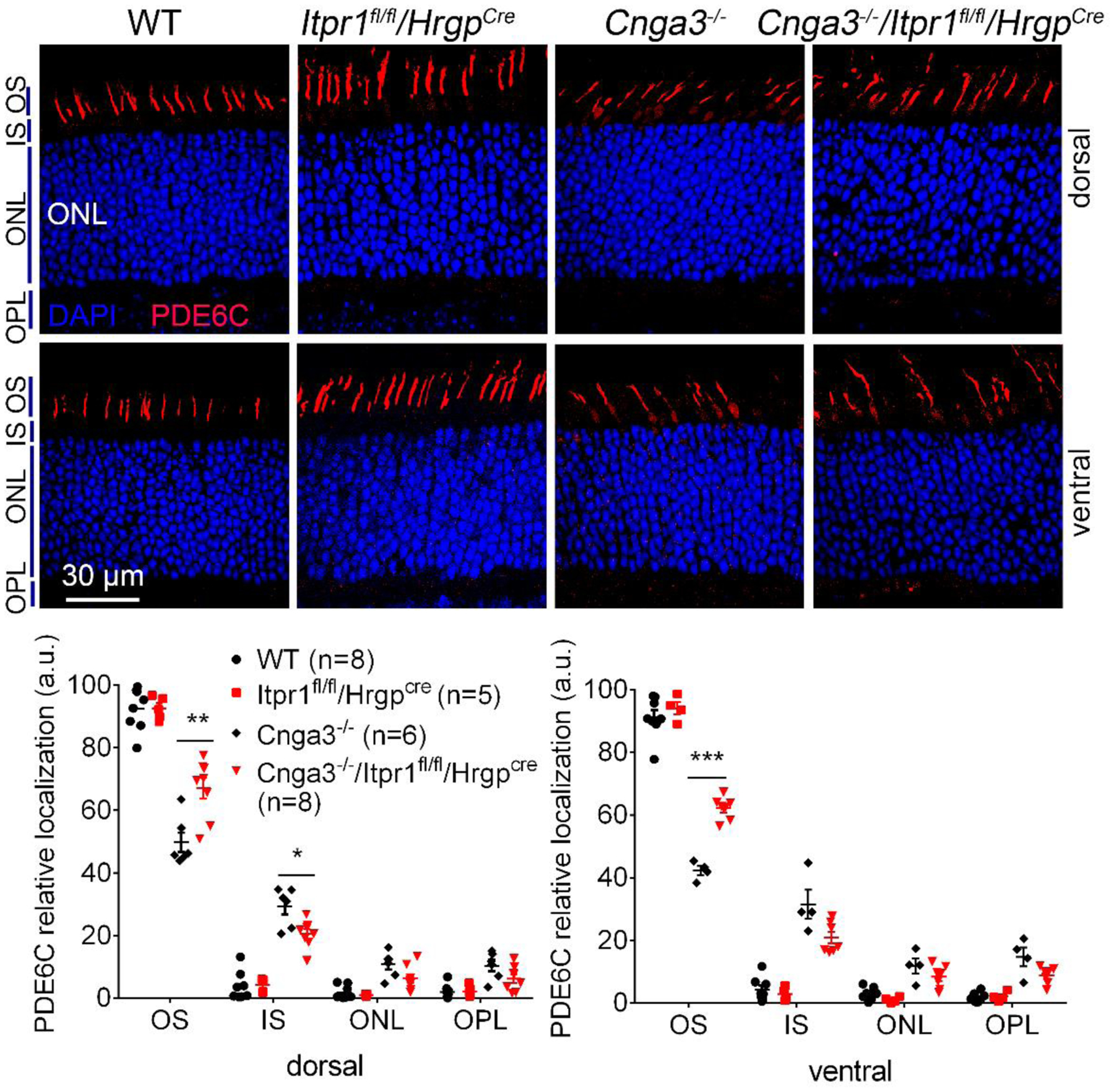
Figure 6. Deletion of IP3R1 increased OS localization of PDE6C in CNG channel-deficient retinas.

PDE6C localization was evaluated by immunofluorescence labeling on retinal cross sections prepared from mice at P30. Shown are representative confocal images of immunofluorescence labeling of PDE6C (A) and corresponding quantitative analysis of immunofluorescence intensity level at different regions of the retinal cross sections (B). OS, outer segment; IS, inner segment; ONL, outer nuclear layer; OPL, outer plexiform layer. Data are presented as mean ± SEM of 3–4 independent assays using retinas/eye sections from 5–8 mice (*, p < 0.05; **, p < 0.01; ***, p < 0.001).
