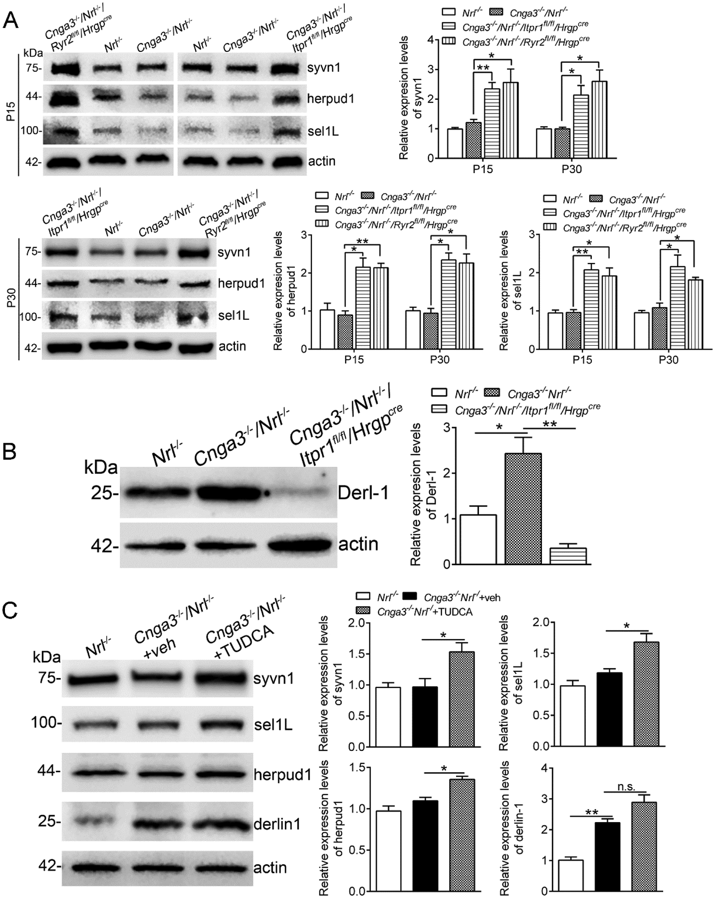
Figure 7. Deletion of IP3R1 or treatment with TUDCA increased expression of ER retrotranslocation proteins in CNG channel-deficient retinas.

Expression levels of ER retrotranslocation proteins in mouse retinas at P15 and P30 (A-B), and after TUDCA treatment (C) were analyzed by immunoblotting. Shown are representative immunoblotting images of these detections and corresponding quantitative analysis, following normalization to internal loading control β-actin. Data are presented as mean ± SEM of 3–4 independent assays using retinas from 8–10 mice (*p < 0.05, **p < 0.01).
