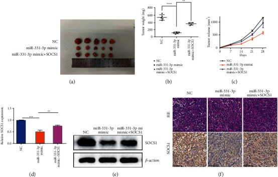
Figure 6.

SOCS1 acts as a target of miR-331-3p to promote tumorigenesis in vivo. 143b cells are inoculated subcutaneously in female nude mice at a density of 5 × 10⁶ cells. (a) After 4 weeks, the tumor is dissected and imaged. (b) Average tumor weight of mice. The data represents the mean ± standard error of the mean (SEM) (n = 5 per group). (c) Tumor volume (ab2/2) was recorded every seven days after mice were injected with stable OS cells. The data represents the mean ± SEM (n = 5 per group). (d) qRT-PCR analysis of SOCS1 mRNA expression in xenograft mouse tumors. (n = 5 in each group). (e) Western blot analysis of SOCS1 protein expression levels in different groups of tumors. (f) HE and immunohistochemistry staining shows the tumor structure and relative protein level of SOCS1 in tumors. The results are shown as the mean ± standard deviation (SD) from three independent experiments. ∗∗P < 0.01.
