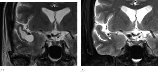
Fig. 2.
Comparison of coronal MRI images before and after the resolution of the cystic lesions
Fig. 2a: Coronal T2 weighted image at the time of initial diagnosis showed the branch of the middle cerebral artery (M2) runs above the cyst.
Fig. 2b: Coronal T2 weighted image showed the cyst had disappeared.

