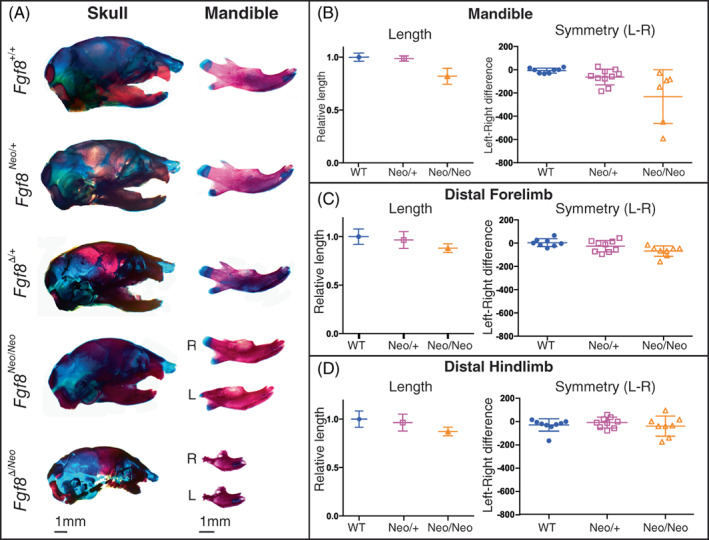
FIGURE 1.

Mandibles exhibit dosage sensitivity and directional asymmetry. (A) Neonatal skulls (left) and isolated dentary bones (right) differentially stained for bone (red) and cartilage (blue). Representative images are shown for Fgf8 +/+ (WT), Fgf8 Neo/+ , Fgf8 Δ/+ , Fgf8 Neo/Neo , and Fgf8 Δ/Neo neonates. Right (R) and left (L) sides of the dentary are shown for the two mutant genotypes. (B) The length (left) of isolated dentaries were measured using arbitrary units and symmetry (right) was determined by subtracting the length of the right side from the length of the left side for each individual. (C, D) Isolated limb bones from Fgf8 Neo/+ and Fgf8 Neo/Neo neonatal mice were measured for length (left) and symmetry (right). n = 9 per genotype. 1 mm scale bars are shown for each respective column in (A)
