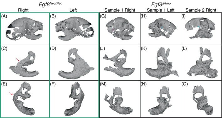
FIGURE 2.

Fgf8‐mediated jaw fusion occurs in two major forms. Three‐dimensional reconstructions from micro‐CTs of (A‐F) Fgf8 Neo/Neo (green box) and (G‐O) Fgf8 Δ/Neo (black box) neonatal skulls and jaws. Panels (C‐F) and (J‐O) show individual outer, then frontal, aspects of a single dentary of the corresponding skull (shown in the upper panels). (A‐F) Fusion of Fgf8 Neo/Neo jaws occurs between a broadened zygomatic arch at the lateral side of the dentary and is more severe on the left side than the right side [arrows in (C) and (E) point to unfused zygomatic contact). (G‐O) Fgf8 Δ/Neo jaws exhibit an additional fusion between the dentary and maxillary process. The zygomatic fusion in Fgf8 Δ/Neo jaws occurs as a thinner bony element that is more distal and lateral on the dentary than what is observed in Fgf8 Neo/Neo jaws. Although the overall fusion is similar on both sides of the jaw in Fgf8 Δ/Neo , the squamosal process can be (G) present (purple asterisk) or (H, I) absent (red askterisks). Data shown are representative of eight Fgf8 Neo/Neo and 7 Fgf8 Δ/Neo neonatal skulls. CT, computed tomography
