FIGURE 3.

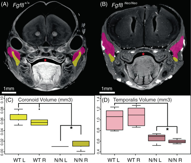
Fgf8 Neo/Neo mutants have small temporalis muscles and absent coronoid processes. Cross‐sections from micro‐CT scans of (A) Fgf8 +/+ (WT) and (B) Fgf8 Neo/Neo neonatal skulls highlighting the coronoid (yellow) and temporalis muscles (pink). (C) Coronoid volume and (D) temporalis volume are represented in mm3. The section where the coronoid was the largest was chosen for each sample in (A, B). The temporalis appears larger in this particular section of the Fgf8 Neo/Neo embryo because shape changes in the muscle related to the smaller jaw and proximal shift in the location of the coronoid relative to the rest of the skull condense the temporalis. Red asterisks highlight normal palate formation in WT and the cleft palate in Fgf8 Neo/Neo . 1 mm scale bars are shown for each panel. *P‐value <.05, Tukey's HSD Test. n = 6 for WT, n = 3 for Fgf8 Neo/Neo . CT, computed tomography
