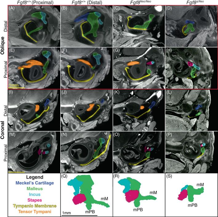
FIGURE 5.

Fgf8 reductions condense the malleus and middle ear. Micro‐CT scans of the middle ear of Fgf8 +/+ (WT; two left columns), Fgf8 Neo/Neo (third column) and Fgf8 Δ/Neo (fourth right column) neonatal skulls. WT sections in the first column are proximal to WT sections in column 2. Rows 1 and 2 (red rectangle) are oblique sections through the malleus and rows 3 and 4 are coronal sections through similar regions. Distal sections are shown in rows 1 and 3; more proximal sections of each sample are shown in rows 2 and 4. (A, B) In WT, the manubrium of the malleus (green) is shown where it contacts the tympanic membrane (TM; yellow) creating a V shape at the point of contact. (C, D) In, Fgf8 Neo/Neo and Fgf8 Δ/Neo the manubrium is dorsally rotated away from the TM, which is small and dysmorphic. (E, F) In WT, the tensor tympani muscle (orange) curves around the sphenoid‐temporal articulation. The tensor tympani muscle is reduced in (G) Fgf8 Neo/Neo , and possibly absent in (H) Fgf8 Δ/Neo [see also (K, L)]. (I, J) In WT, the tympanic cavity is large separating the middle ear and cochlea, but in (K) Fgf8 Neo/Neo the cavity is reduced in size and becomes smaller in (L) Fgf8 Δ/Neo . (M, N) The incus (light blue) articulates with the stapes (pink) at the oval window of the otic capsule proximal to the malleus [panel (N) is distal to (M)]. (O, P) In the mutants, the middle ear is compressed, such that all three middle ear bones lie in one plane, with the incus and stapes rostral to the malleus. The stapes appears morphologically normal in all dosage levels. (Q‐S) Three‐dimensional reconstructions of the middle ear of (Q) Fgf8 +/+ (WT), (R) Fgf8 Neo/Neo , and (S) Fgf8 Δ/Neo ; malleus, incus and stapes. Manubrium of Malleus (mM),process brevis of the malleus (mPB). Scale bar in (E) applies to (A‐H), scale bar in (M) applies to (I‐P), and scale bar in (Q) applies to (Q, S). CT, computed tomography
