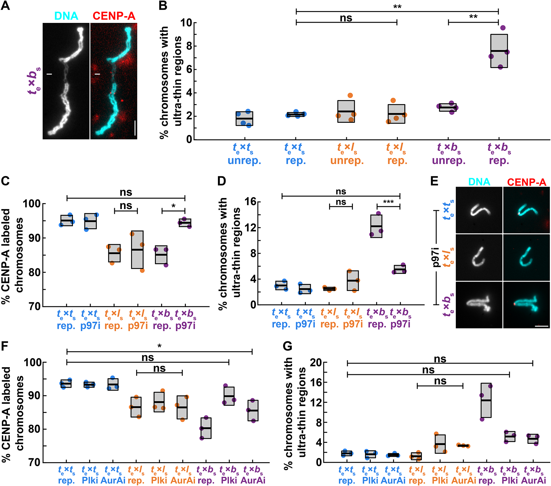
Figure 4: Mitotic replication stress leads to X. borealis centromere and chromosome morphology defects.

(A) Representative image showing an ultra-thin region of a mitotic X. borealis chromosome formed in X. tropicalis egg extract. Note that the chromosome has an intact centromere. DNA in cyan, CENP-A in red. Scale bar is 5 μm.
(B) Percentage of unreplicated and replicated mitotic chromosomes with ultrathin morphology defects in X. tropicalis extract. A low percentage of X. tropicalis, X. laevis or X. borealis unreplicated chromosomes display ultra-thin regions. After cycling through interphase, only X. borealis chromosomes exhibit a significant increase in this defect. Quantification with N = 3 extracts, N > 310 chromosomes per extract. p-values (top to bottom, then left to right) by one-way ANOVA with Tukey post-hoc analysis: 2.9352e-7, 0.9999, 1.6475e-6.
(C) Percentage of replicated chromosomes with centromeric CENP-A staining in X. tropicalis extracts treated with solvent control or 10 μM p97 ATPase inhibitor NMS-873 (p97i). Inhibition of p97 restores CENP-A staining on X. borealis mitotic chromosomes, but does not affect X. tropicalis or X. laevis chromosomes. p-values (top to bottom, then left to right) by one-way ANOVA with Tukey post-hoc analysis: 0.9997, 0.9978, 0.0204.
(D) Percentage of chromosomes with ultrathin regions in X. tropicalis extracts treated with solvent control or 10 μM p97 ATPase inhibitor NMS-873 (p97i). Inhibition of p97 rescues X. borealis chromosome morphology defects, but does not affect X. tropicalis or X. laevis chromosomes. p-values (top to bottom, then left to right) by one-way ANOVA with Tukey post-hoc analysis: 0.1114, 0.6903, 6.2572e-5.
(E) Representative images of mitotic replicated X. tropicalis, X. laevis, and X. borealis chromosomes following treatment with 10 μM p97 ATPase inhibitor NMS-873 (p97i). X. borealis chromosome morphology and centromere localization are rescued (bottom panels, compare to Fig. 4A, 2B images), similar to X. tropicalis, while X. laevis chromosomes have lost CENP-A staining (middle panels). DNA in cyan, CENP-A in red. Scale bar is 5 μm.
(F) Percentage of replicated chromosomes with centromeric CENP-A staining in X. tropicalis extracts treated with solvent control, 1 μM Polo-like kinase 1 inhibitor BI-2536 (Plk1i), or 1 μM Aurora A kinase inhibitor MLN-8237 (AurAi). CENP-A localization is fully or partially rescued on X. borealis mitotic chromosomes, whereas X. tropicalis or X. laevis chromosomes are not affected. p-values (top to bottom) by one-way ANOVA with Tukey post-hoc analysis: 0.0276, 0.7003, 0.9999.
(G) Percentage of chromosomes with ultrathin regions in X. tropicalis extracts treated with solvent control, 1 μM Polo-like kinase 1 inhibitor BI-2536 (Plk1i), or 1 μM Aurora A kinase inhibitor MLN-8237 (AurAi). Inhibition of Plk1 and AurA rescued X. borealis mitotic chromosome morphology defects, but did not affect X. tropicalis or X. laevis chromosomes. p-values (top to bottom) by one-way ANOVA with Tukey post-hoc analysis: 0.2882, 0.1525, 0.5887.
C, D: N = 3 extracts, N > 179 chromosomes per extract.
E, F: N = 3 extracts, N > 155 chromosomes per extract.
B-F: ns, not significant.
See also Figure S4.
