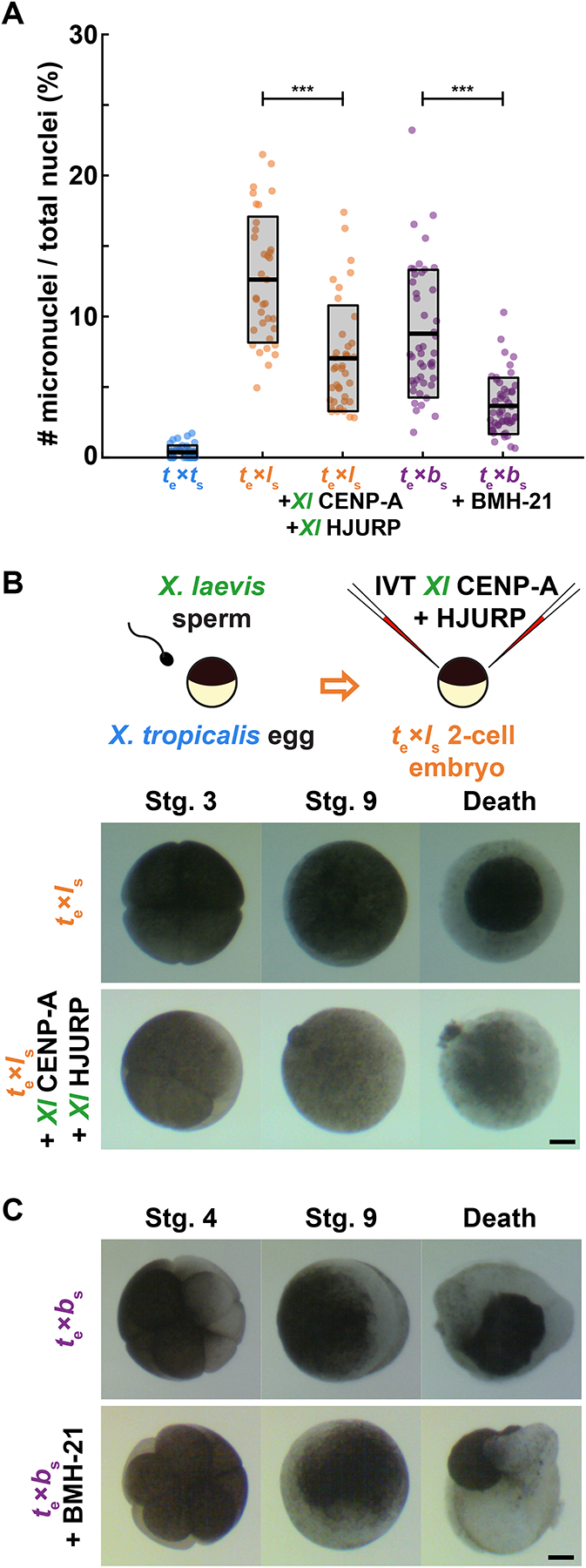
Figure 6: Treatments that rescue CENP-A localization in egg extracts reduce micronuclei formation in hybrid embryos, but inviability persists.

(A) Quantification of chromosome mis-segregation events as measured by the number of micronuclei compared to total nuclei in treated hybrid embryos. X. tropicalis eggs fertilized with X. laevis sperm were microinjected with X. laevis CENP-A/HJURP, while X. tropicalis eggs fertilized with X. borealis sperm were treated with Pol 1 inhibitor BMH-21. Embryos were fixed at stage 9 (7 hpf) just before gastrulation and hybrid death. The number of micronuclei was significantly reduced in both cases, but not to control levels measured in X. tropicalis eggs fertilized with X. tropicalis sperm. N = 3 clutches for each hybrid, N > 15 embryos and > 200 cells per embryo. p-values (left to right) by two-tailed two-sample unequal variance t-tests: 2.111e-7, 2.651e-9; ns, not significant.
(B) Schematic of experiment and video frames of X. tropicalis eggs fertilized with X. laevis sperm microinjected at the two-cell stage with X. laevis CENP-A/HJURP, increasing centromeric protein concentration by ~44.5%. Microinjected hybrid embryos die at the same time and in the same manner as uninjected hybrid controls. N = 10 embryos across 4 clutches. Scale bar is 200 μm. See also Video S1.
(C) Video frames of X. tropicalis eggs fertilized with X. borealis sperm that were incubated from the two-cell stage with 1 μM RNA Pol I inhibitor, BMH-21. Treated hybrid embryos die at the same time and in the same manner as untreated hybrid controls. N = 12 embryos across 2 clutches. Scale bar is 200 μm. See also Video S2.
See also Figure S6.
