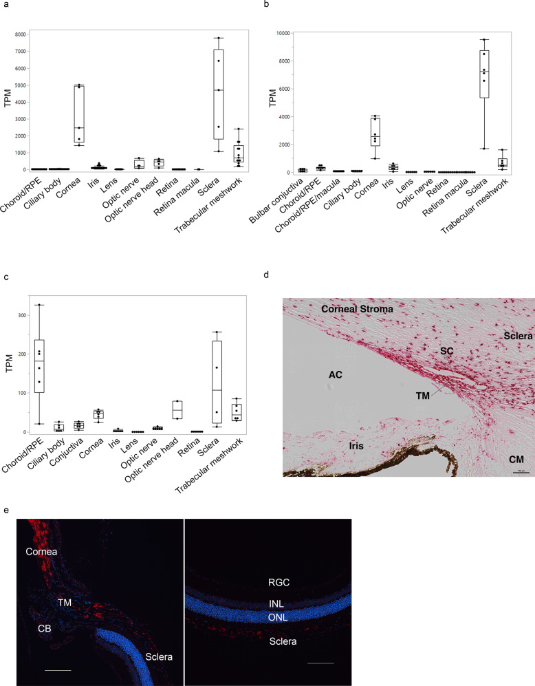
Fig. 5. ANGPTL7 expression in ocular tissues across species.
RNA-sequencing-based expression levels (measured in transcripts per million, TPM, and represented as median and interquartile range) are highest in cornea, trabecular meshwork (TM), and sclera in human (a), and African green monkey (b) eyes, and in cornea, TM, sclera, optic nerve head, and choroid/RPE in C57BL/6 J mouse eyes (c); In situ hybridization (RNAscope) shows ANGPTL7/Angptl7 (red) expression in TM, cornea and sclera in human (d) and murine (e) eyes. Scale bars represent 100 μm. DAPI staining (blue) counterstains cell nuclei. RPE retinal pigmented epithelium, CB ciliary body, SC Schlemm’s canal, CM ciliary muscle, AC anterior chamber, RGC retinal ganglion cell, INL inner nuclear layer, ONL outer nuclear layer.

