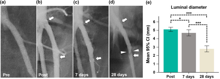
Figure 2.
Representative follow-up angiographies with the mean luminal diameters in the stented LIAs. (a) Pre-angiography shows the LIA. (b) Post-angiography shows the fully expanded TPU and GL blended NFs-covered stent-graft (arrows). (c) Follow-up angiography at 7 days shows good patency of the stent-graft (arrows). (d) Follow-up angiography at 28 days shows an irregular luminal narrowing (arrowheads) at the distal portion of the TPU and GL blended NFs-covered stent-graft (arrows). (e) The graph shows changes in the luminal diameters in the stented LIAs. Note. CI, confidence interval; Note. TPU: thermoplastic polyurethane, GL: gelatin, NF: nanofiber; *p < 0.05, ***p < 0.001.

