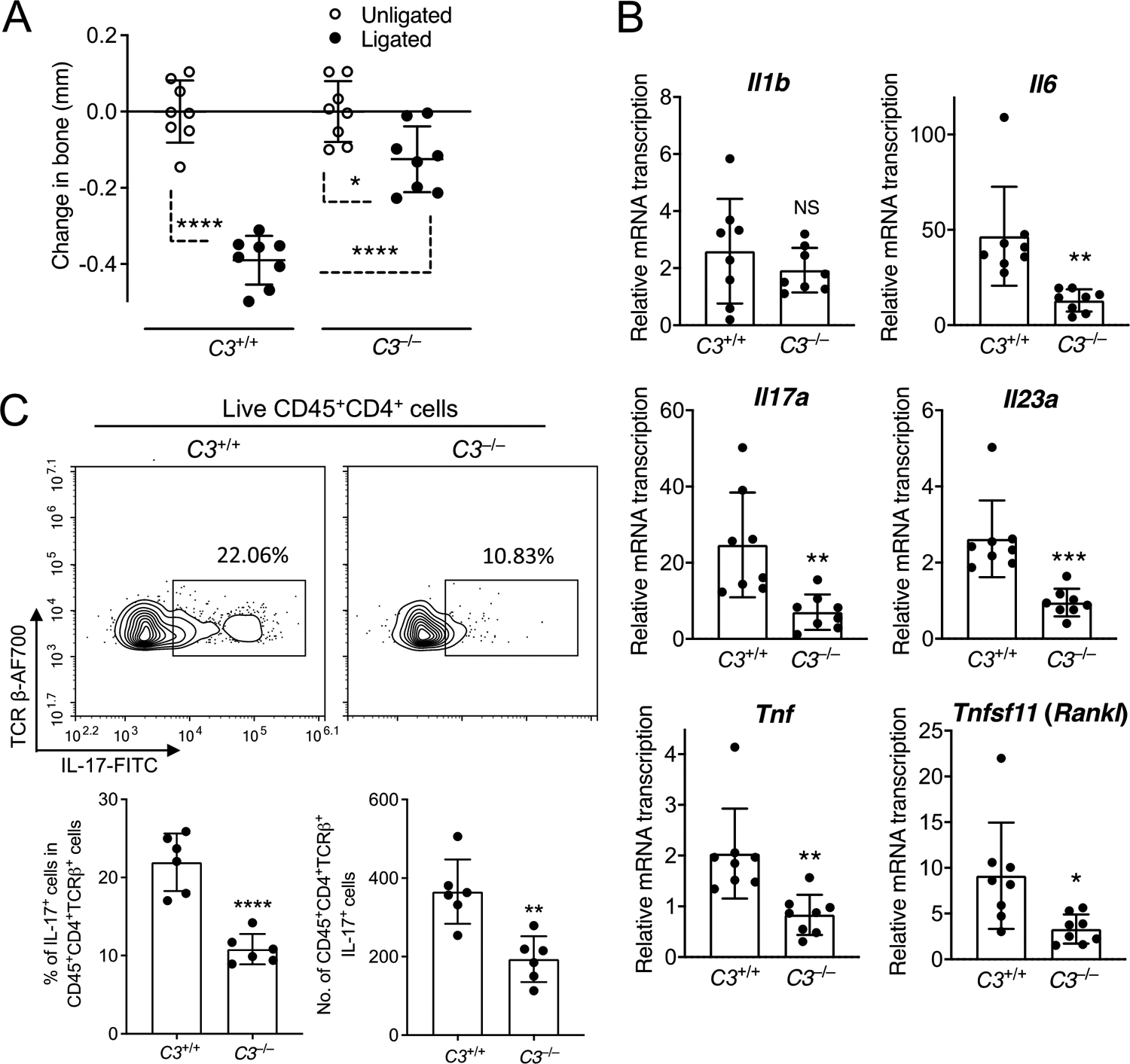
Figure 2: C3 deficiency inhibits LIP-induced periodontal inflammation and Th17 cell accumulation.

C3–/– mice and C3+/+ littermate controls were subjected to ligature-induced periodontitis (LIP) for 5 days by ligating a maxillary second molar and leaving the contralateral tooth unligated to serve as baseline control. (A) Bone loss determination relative to the unligated baseline. (B) Relative gingival mRNA expression of indicated cytokines determined by quantitative real-time PCR. (C) Representative FACS plots of Th17 cells in gingival tissue (top) and bar graphs showing percentage of Th17 cells in CD4+ T cells (bottom left) and Th17 absolute numbers (bottom right) in the gingival tissue of C3–/– mice and C3+/+ controls on day 5 after LIP. Data are means ± SD (A,B: n = 8 mice per group; C: n = 6 mice per group). *P < 0.05, **P < 0.01, ***P < 0.001, ****P < 0.0001 between indicated groups (A: One-way ANOVA with Tukey’s multiple comparison test; B,C: Unpaired Student’s t-test). NS, not significant.
