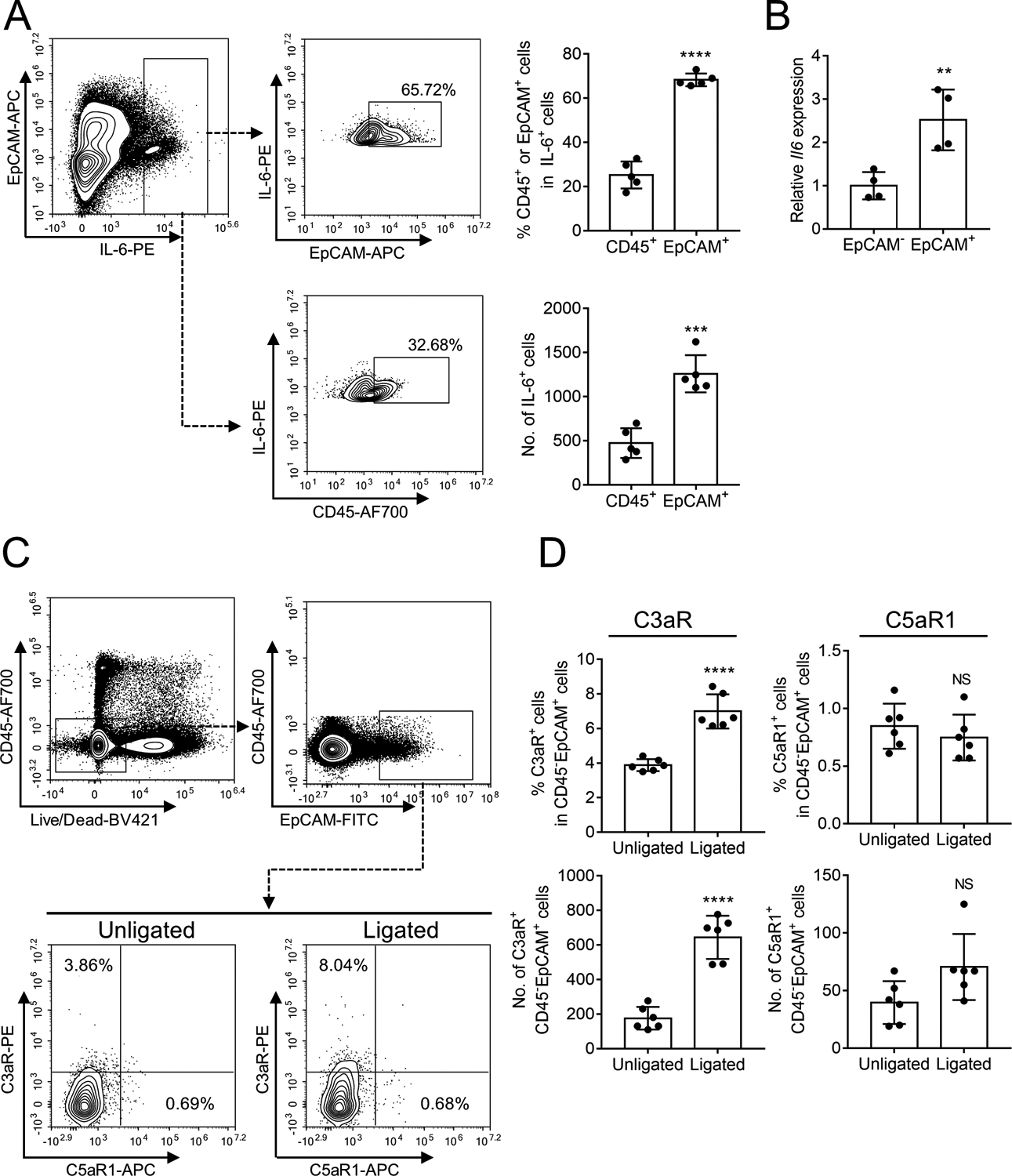
Figure 4. LIP-induced IL-6 is predominantly produced by epithelial cells that also express C3aR.

(A-B) Gingival tissues were collected from ligated sites of ligature-induced periodontitis (LIP)-subjected mice and processed for FACS analysis to detect IL-6-expressing leukocytes (CD45+) or epithelial cells (EpCAM+). (A) Representative FACS plots (left) and bar graphs showing percentage of IL-6-expressing cells in CD45+ cells and EpCAM+ cells (top right) and absolute numbers of IL-6+CD45+ cells and IL-6+EpCAM+ cells (bottom right) in the gingival tissue on day 5 after LIP. (B) Gingival cells were sorted into EpCAM+ and EpCAM– cells and analyzed for IL-6 mRNA expression by quantitative real-time PCR. (C,D) Gingival tissues were collected from LIP-subjected mice and processed for FACS analysis to detect expression of C3aR and C5aR1 in CD45−EpCAM+ cells. (C) Representative FACS plots and (D) bar graphs showing percentage of C3aR-expressing or C5aR1-expressing cells in CD45−EpCAM+ cells (top) and absolute numbers of C3aR+CD45−EpCAM+ cells or C5aR1+CD45−EpCAM+ cells (bottom) in unligated and ligated sites on day 5 after LIP. Data are means ± SD (n = 4–6 mice per group). **P < 0.01, ***P < 0.001, ****P < 0.0001 between indicated groups (Unpaired Student’s t-test). NS, not significant.
