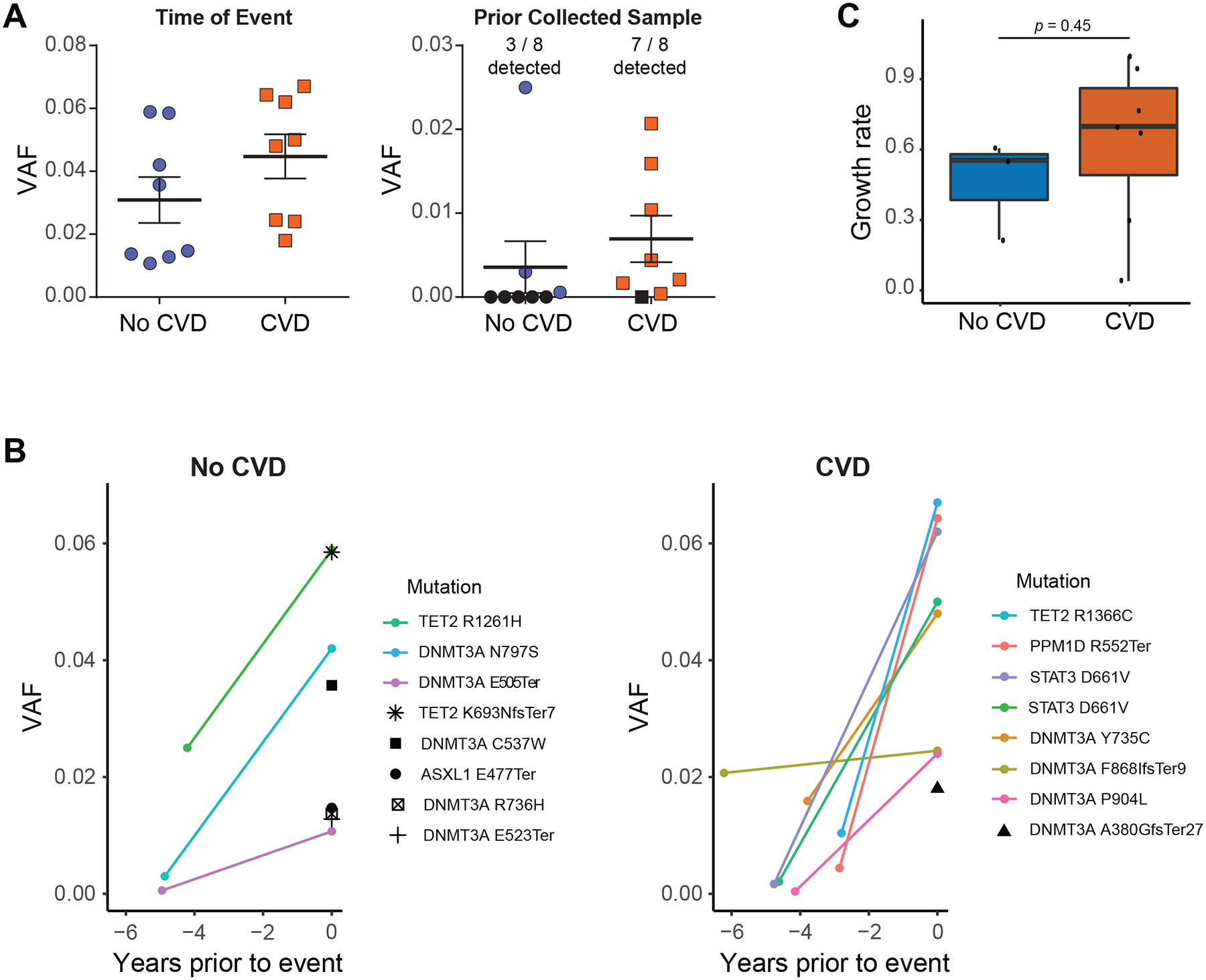
Figure 2. CH is a Biomarker for CVD in PLWH.

(A) VAF of CH mutations in PBMCs from PLWH quantified by ddPCR at the time of CVD event or a prior collected sample. The CH mutation identified in CVD cases at the time of event was observed in the prior collected sample for 7 out of 8 patients, whereas only 3 out of 8 CH clones were identified in the prior banked sample for PLWH without CVD. Black shapes denote a mutation was not detected. (B) Change in VAF of mutations between the initial blood collection timepoint and the collection closest to the CVD event or last follow-up for cases and controls respectively. Mutations that were detected at both times points are displayed using exponential growth curves while mutations that were only detectable at 2nd blood draw are shown with single black shapes. (C) Calculated clonal expansion rate for CH mutations among PLWH with and without CVD. Shaded bands represent intra-quartile ranges.
