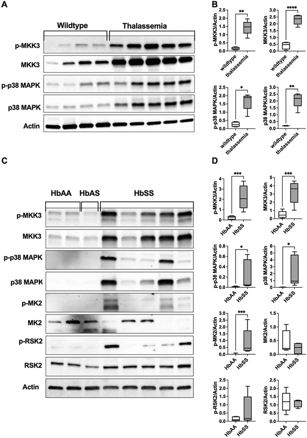
Fig. 6.
Differential expression of erythrocyte MAPK proteins in thalassemia and sickle cell disease. Western blotting analyses of erythrocyte membranes from a mouse model of β-thalassemia (B6.129P2-Hbb-b1tm1Unc Hbb-b2tm1Unc/J) or from patients with sickle cell disease. A. Representative immunoblots of phosphorylated (p-) and total MKK3, p38 MAPK, and actin in erythrocyte ghost from thalassemic mice and wildtype controls (C57BL/6J). B. Quantified protein expressions (normalized to actin) are shown for phospho-MKK3, total MKK3, phospho-p38 MAPK, and total p38 MAPK. N = 5 (thalassemic mice) and N = 4 (wildtypes). *p = 0.0159 by Mann–Whitney test; **p < 0.002 and ****p < 0.0001 by unpaired t-test. C. Representative immunoblots of phosphorylated (p-) and total MKK3, p38 MAPK, MAPAPK2 (MK2), RSK2, and actin in erythrocyte membranes from healthy volunteers (HbAA), a male donor with sickle cell trait (HbAS), and patients with sickle cell disease (HbSS). D. Quantified protein expressions (normalized to actin) are shown for phospho-MKK3, total MKK3, phospho-p38 MAPK, total p38 MAPK, phospho-MAPAPK2 (MK2), total MK2, phospho-RSK2, and total RSK2. N = 5 (HbSS) and N = 8 (HbAA). *p < 0.05, ***p ≤ 0.0008 by unpaired t-test, except for p-MK2 where Mann–Whitney test was used. Data were collected from 2 (panel B) and 3 (Panel D) independent experiments and are presented as box and whiskers (median and min to max).

