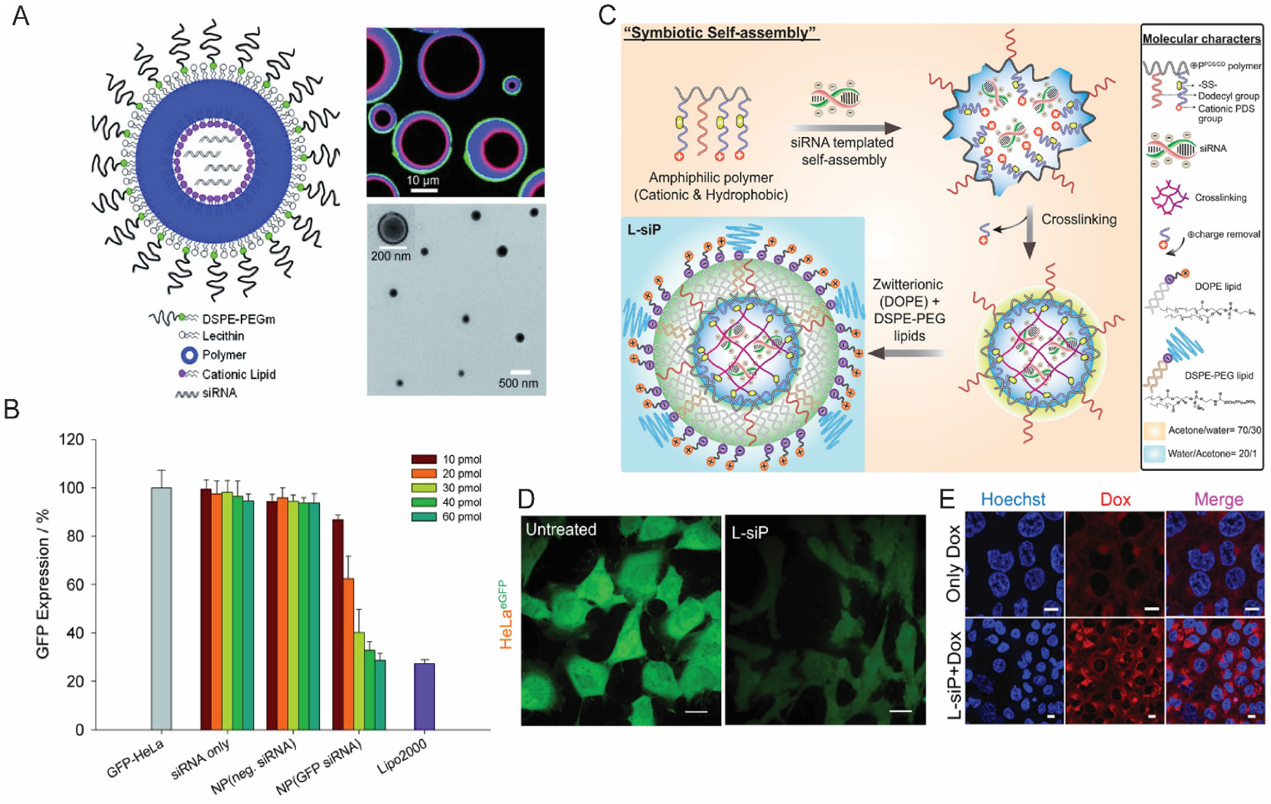
Figure 2:

(A) A schematic illustration of core/shell lipid-polymer-lipid hybrid nanomaterial, with confocal microscopy image (right top) and TEM image (right bottom) confirming the core/shell morphology (B) Flow cytometry data showing dose dependent GFP knockdown in HeLa cells. (Reproduced with permission from reference 54, Copyright 2011 Wiley-VCH Verlag GmbH & Co) (C) A schematic illustration depicting the formulation procedure of “virus mimicking” lipid-polymer hybrid particles. (D) Confocal microscopy data showing efficient knockdown of GFP in HeLa cells; scale bar, 20 μm. (E) Effect of treating NCI-ADR/RES cells with MDR1 siRNA using lipid-polymer hybrid nano-assemblies; scale; 10 μm. (Reproduced with permission from reference 61, Copyright 2019 American Chemical Society)
