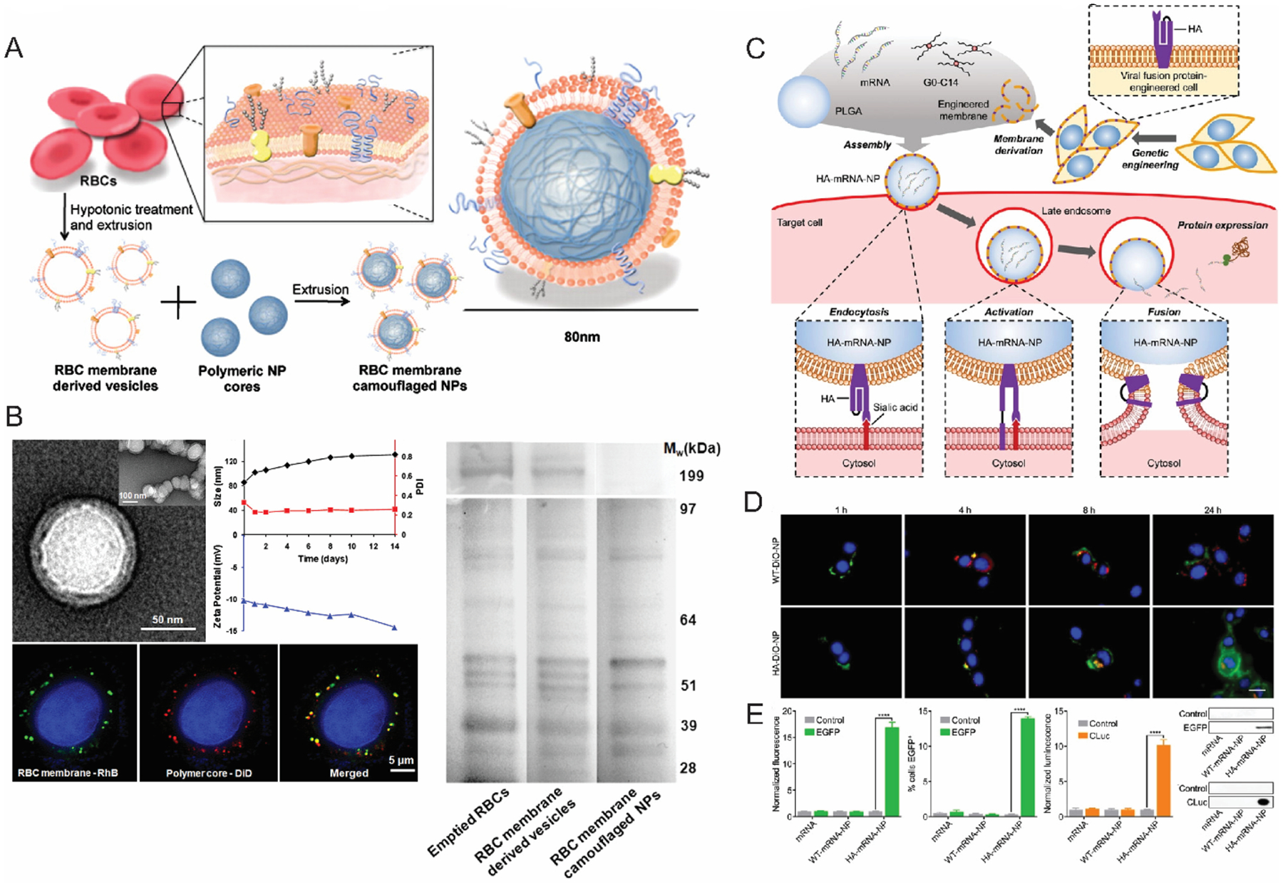
Figure 3:

(A) A schematic illustration of RBC membrane coated PLGA nanoparticles (B) TEM and confocal microscopy characterization of RBC membrane coated nanoparticles (left); size and zeta potential data showing the stability of nanoparticles; SDS-PAGE data (right) shows the similarity of protein expression of the RBC derived membranes with native RBC membranes (Reproduced with permission from reference 65, Copyright 2011 The National Academy of Sciences of the USA) (C) mRNA delivery strategy utilizing genetically modified cells expressing viral fusion protein hemagglutinin (HA). (D) Endosomal escape and cytosolic localization of Dio-labeled nanoparticles in B16-WT cells; scale bar 20 μm (E) Expression of eGFP and cLUC in B16-WT cells, confirmed with flow cytometry and western blots. (Reproduced with permission from reference 83, Copyright 2022 Wiley-VCH Verlag GmbH & Co)
