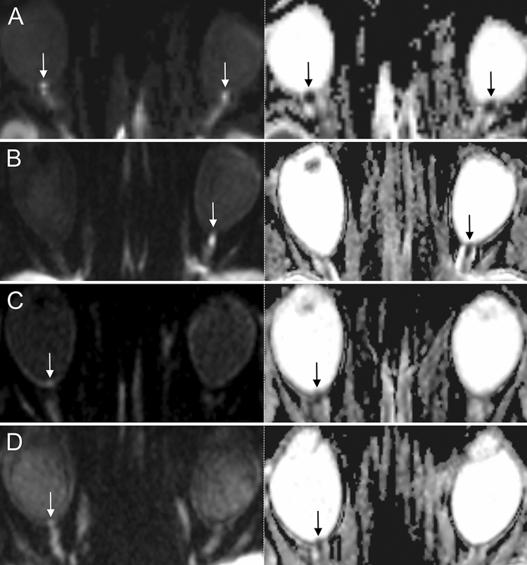
Figure 2.
Examples of 3 T DWI-MRI in 4 GCA patients presenting with acute arteritic AION (left row: DWI b = 1000 s/mm2, right row: corresponding ADC images). (A, B): Distinct findings of ONH restricted diffusion in 2 patients with AION (white arrows; bilateral (A) and left-sided (B)). Note the marked qualitative ADC reduction in both cases (black arrows). (C, D) Subtle findings of ONH restricted diffusion (white arrows) and corresponding visually qualitative ADC reduction (black arrows) in 2 patients with right-sided AION. Abbreviations: AION, anterior ischemic optic neuropathy; DWI-MRI, diffusion-weighted magnetic resonance imaging; GCA, giant cell arteritis; ONH, optic nerve head.

