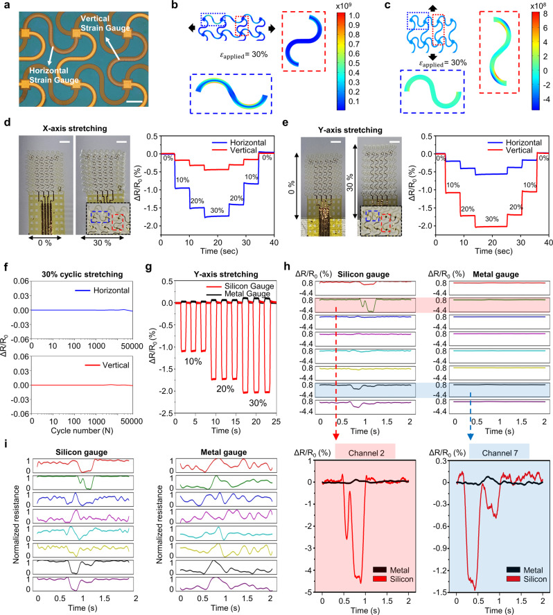
Fig. 2. Characterizations of SiNM-based biaxial strain gauge.
a Magnified optical image of biaxial strain gauges comprising a horizontal gauge and a vertical gauge; scale bar, 300 µm. b, c Finite element analysis of applied local strain to the biaxial strain gauge on an elastomer substrate with 30% stretching along x (b) and y (c) directions. d, e Photographs of the biaxial strain gauges before and after 30% stretching test (left) and resistance change of both horizontal and vertical gauges during in vitro test at 10%, 20%, and 30% stretching (right) in x (d) and y (e) directions, showing independent sensing properties of the biaxial strain gauges where the stretching in parallel and perpendicular directions to the gauge is prone to apply dominant and minor strain, respectively; scale bars, 1 mm. Inset: enlarged photos of a biaxial strain gauge under an applied strain. f Relative change in electrical resistance during 50000 cycles of 30% stretching along y direction under 10 mm/s. Each cycle has a start delay and end delay of 1 s. g Comparison of sensitivity to strain between SiNM- and metal-based strain gauge through 10%, 20%, and 30% cyclic stretching test. h Waveforms of corresponding in vivo test of both SiNM- and metal-based strain gauges during silent speech of the word, WITHOUT (h, top), and magnified plots of channels 2 (red highlight) and 7 (blue highlight) (h, bottom). i Normalized waveforms of h in the training phase.

