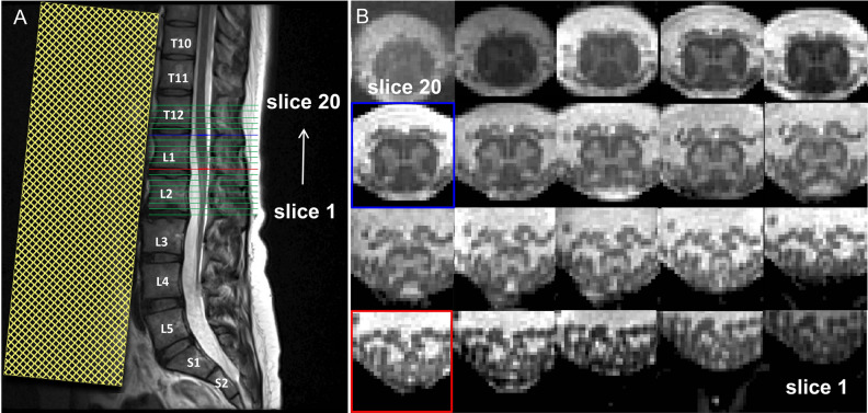
Figure 1.
(A) Sagittal T2-weighted turbo spin echo acquisition in the lower spine used for subsequent prescription of the high-resolution axial acquisition. (B) Corresponding axial slices acquired with the 3D multi-echo gradient-echo sequence ( Siemens FLASH) in the caudal-rostral direction (slices 1–20). Highlighted are the slice in the lumbosacral enlargement (LSE) with the largest cord cross-sectional spinal cord area (defined as the "LSE slice" and shown in light blue in A and B; here: slice 15), and the most caudal slice in the conus medullaris (CM) where the gray matter still has the characteristic butterfly shape (defined as the "CM slice" and shown in red in A and B; here: slice 9). A saturation band, displayed as yellow shaded area in A, was placed anterior to the spine to suppress signal and possible artifacts arising from abdominal peristalsis.

