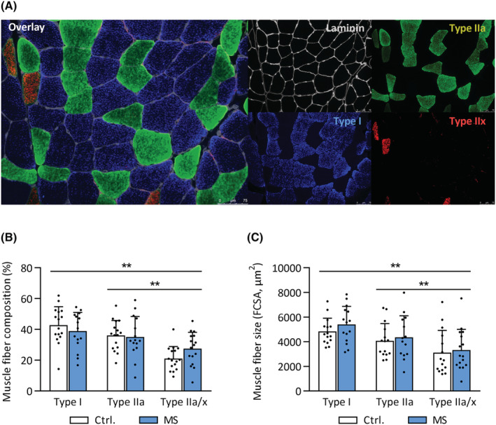
Figure 2.

Normal muscle fibre size and composition in MS. Vastus lateralis biopsies were obtained from MS patients and healthy controls. (A) Representative immunohistochemistry images showing the presence of myosin heavy chain type I, IIa, and IIx, delineated by laminin‐stained cell membranes. Scale bar: 75 μm. (B) Muscle fibre type composition of type I, IIa, and IIa/x fibres in MS and controls. (C) Fibre cross‐sectional area (FCSA) of type I, IIa, and IIa/x fibres in MS and controls. **P < 0.01 for main effect of fibre type (from mixed model analysis). Mean ± SD.
