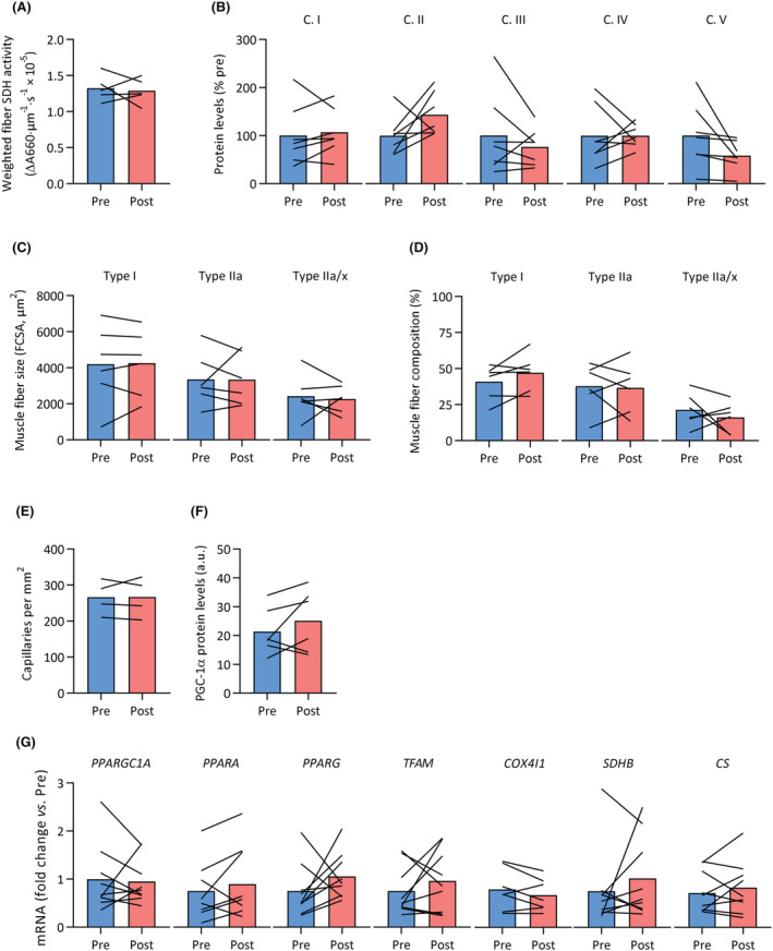
Figure 7.

Skeletal muscle fibre composition, fibre size, oxidative capacity, mitochondrial OXPHOS protein levels, capillarity and mitochondrial signalling did not respond to exercise training in MS. Vastus lateralis biopsies were obtained from patients with MS before (pre, blue) and after (post, red) a 12 week exercise training intervention. No significant differences were observed in (A) SDH activity weighted for muscle fibre type, (B) mitochondrial OXPHOS protein levels (complex [C.] I to V), (C) fibre cross‐sectional area (FCSA) for type I, IIa and IIa/x fibres, (D) fibre type composition, (E) capillary density, (F) PGC‐1α protein content, and (G) mRNA expression of various mitochondrial transcription factors and genes.
