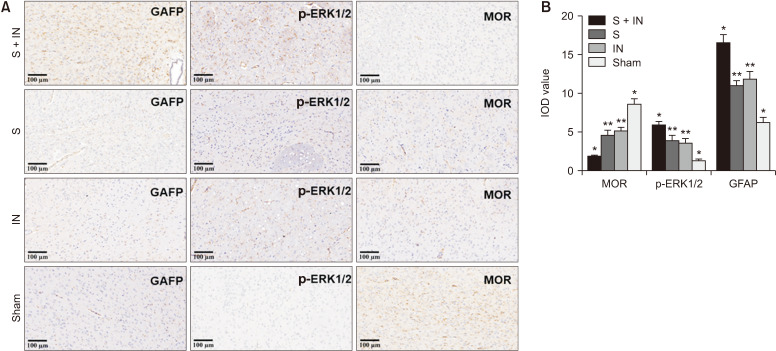
Fig. 4.
(A) The expression of GFAP, p-ERK1/2, and MOR by immunohistochemistry staining in amygdala tissue of rats (magnification: 200×). The positive expressing proteins appeared brown in color. (B) The results showed that GFAP and p-ERK1/2 are significantly up-regulated in group S + IN (*P < 0.050), the expression of MOR is significantly down-regulated in group S + IN (*P < 0.050). There were no significant differences between group IN and group S (**P > 0.050). The expression of GFAP and p-ERK1/2 are significantly up-regulated in group S and group IN (*P < 0.050) compared with the sham group. The expression of MOR is significantly down-regulated in group S and group IN (*P < 0.050) compared with the sham group. The error bars indicate mean ± standard deviation. GFAP: glial fibrillary acidic protein, p-ERK1/2: phosphorylated extracellular regulatory protein kinase, MOR: mu-opioid receptors, S: stress, IN: incision, IOD: integrated optical density.

