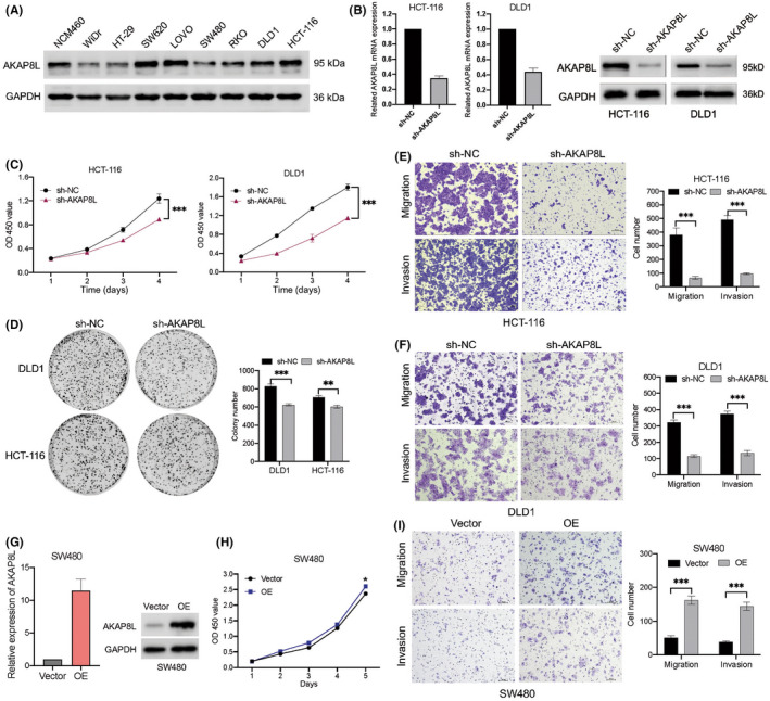
FIGURE 9.

AKAP8L promotes colon cancer cell growth, migration, and invasion. (A) Basic protein expression of AKAP8L in normal colonic epithelial cell lines and colon cancer cell lines detected by western blot. GAPDH serve as the loading control. (B) mRNA and protein levels of AKAP8L in HCT‐116 and DLD1 cells with control vector (sh‐NC) or sh‐AKAP8L were evaluated by quantitative PCR and western blot. (C) Cell growth in HCT‐116 and DLD1 cells with sh‐NC or sh‐AKAP8L was determined by CCK‐8 assay. (D) Left, representative images of cell colonies stained with crystal violet in HCT‐116 and DLD1 cells with sh‐NC or sh‐AKAP8L. Right, numbers of colonies as mean ± SD from three independent experiments. (E,F) Left, representative images of migrated cells in migration and invasion assays were stained with crystal violet in HCT‐116 cells with sh‐NC or sh‐AKAP8L. Right, numbers of cells as mean ± SD from three independent experiments. (G) mRNA and protein levels of AKAP8L in SW480 cells transfected with sh‐NC or AKAP8L overexpression (OE) vector. (H) Cell growth in SW480 cells with vector or AKAP8L‐OE determined by CCK‐8 assay. (I) Representative images and statistic graph of migrated cells in migration and invasion assays in in SW480 cells with vector or AKAP8L‐OE. Data are presented as mean ± SD. **p < 0.01, ***p < 0.001 (Student's t‐test). OD, optical density
