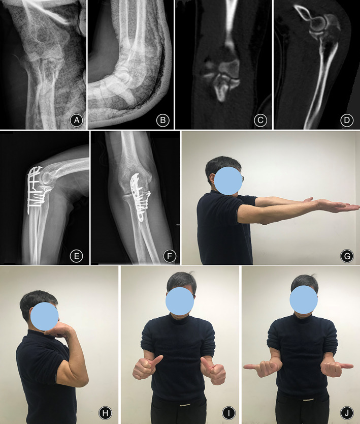
Fig. 3.

A typical case of the O'Driscoll type 1 fracture using suture‐preset spring plate system (SSPS) fixation. The preoperative X‐ray in AP (A) and lateral (B) view showed fracture of the coronoid process tip. (C and D) The fracture was comminuted in CT scan view. The X‐ray in AP (E) and lateral (F) view at 8 months postoperatively. The extension (G), flexion (H), pronation (I) and supination (J) showed good function recovery of the elbow at last follow up
