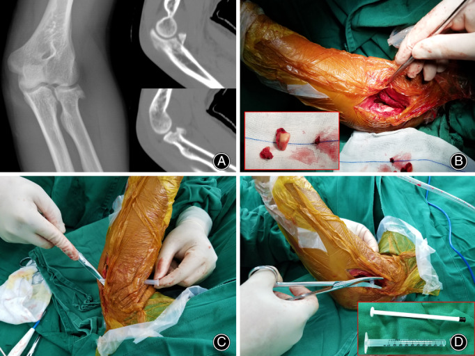
Fig. 1.

Pre‐op and intra‐op findings: Anteroposterior (AP) X‐ray of the radial head fracture and lateral CT scan of the coronoid and radial head fractures (A), view of the coronoid fracture through the lateral incision after removal of the radial head fragment shown in the inset image (B), intra‐operative insertion of the 1 mL syringe guide (C, D).
